2.2 Movement: functions of muscle
Focus now shifts to the function of movement, and the muscle tissue that facilitates it.
All muscles are contractile and excitable and they contain the proteins myosin and actin, which are responsible for contraction. However, the functions of the muscle types and the arrangement of cells within them vary. As you read earlier in the course, from a histological perspective there are three different types of muscle:
- striated (or skeletal) muscle
- smooth muscle
- cardiac muscle.
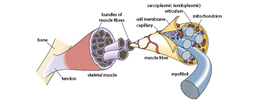
Skeletal muscles are the voluntary muscles, attached to bones via tendons, which allow voluntary movement of the body. Muscle tissue is made from a collection of highly specialised cells, known as muscle fibres. These are formed by the fusion of individual muscle cells (myocytes).
The muscle tissue is surrounded by supporting and protecting bands of fibrous connective tissue called fascia. The plentiful nerve and blood supplies which an active muscle requires are located within fascia.
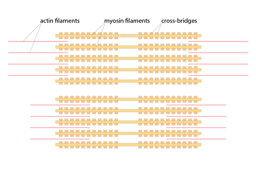
Within a myofibril, actin and myosin are interleaved, producing the characteristic striated (striped) appearance, which can be seen under the microscope and is shown schematically in the figure below. When a muscle contracts the actin and myosin slide across each other, causing the striations to bunch up.
Energy is required to relax the muscle (surprisingly the default condition is contraction). When a muscle runs out of energy it contracts, as occurs in cramp and rigor mortis.

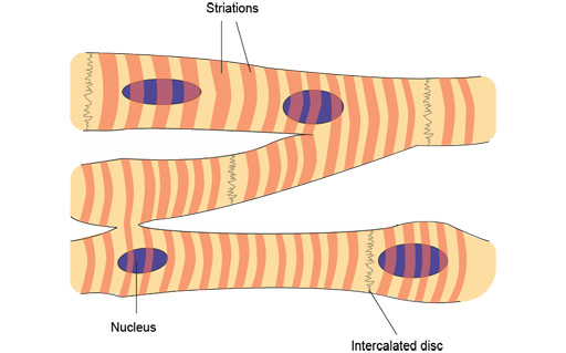
In contrast to skeletal muscle, both cardiac and smooth muscle are involuntary muscles, meaning that we cannot voluntarily control their contraction. In cardiac muscle the myocytes form a network, with crosslinks and intercalated discs between the cells. They have striations comparable to those in skeletal muscle.
Smooth muscle is found in the wall of blood vessels and many hollow organs, including the uterus, the bladder and along all parts of the gastrointestinal tract.
Activity 3
Open the virtual microscope [Tip: hold Ctrl and click a link to open it in a new tab. (Hide tip)] in a new window or tab. Find Slides 4–6 in the ‘Week 3’ category.
See if you can identify the characteristics of the three different types of muscle: striated muscle (Slide 4), cardiac muscle (Slide 5) and smooth muscle (Slide 6).
Note that smooth muscle is illustrated in Slide 6 in the wall of an artery. However, it can also be seen in different areas of the gut (see Slides 3–5 in the ‘Week 2’ category).


