

2.3 Structure and strength: functions of bone and cartilage
Tissues such as bone and cartilage give our bodies structure and strength. The examples used are bone and cartilage.
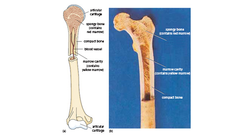
There are many different types of cartilage, including articular cartilage, which covers the joints at the end of the bones. However, the example provided in the slide set that you will examine shortly is from the trachea (windpipe). This structure has bands of hyaline cartilage, which strengthen the trachea, to resist collapse or closure as the head moves.
Notice in these strong tissues how the cells form only a small proportion of the total tissue mass. They are responsible for forming, remodelling and repairing the tissue, but the strength is provided by the extracellular matrix (and mineralisation), including proteins such as collagen and elastin in tendons.
Mineralisation produces highly ordered structures around the cells, and is arranged to resist the stresses placed on the tissue. The hardest tissues of the body – bones and teeth – are mineralised with calcium hydroxyl apatite.
Activity 4
Open the virtual microscope [Tip: hold Ctrl and click a link to open it in a new tab. (Hide tip)] in a new window or tab. Find Slides 7–8 in the ‘Week 3’ category.
First, take a look at the section of bone shown in Slide 7. Identify the cells, osteoblasts (which produce the bone) and osteoclasts (which remodel it), and familiarise yourself with their appearance and structure.
Next, in the section of trachea shown in Slide 8, identify the chondrocytes that form the connective tissues, and in each case relate the position of the cells to the extracellular structures.