2.4 Excretion: functions of the kidney
The kidneys are excretory organs that perform three main functions to produce urine, filtration, reabsorption and secretion. They are situated at the back of the peritoneal cavity.
A cross-sectional slice through the kidney shows an outer layer (renal cortex), a middle layer (renal medulla), and an inner area (renal pelvis), where the ureter widens to join the kidney. These three structures are shown in Figure 10.
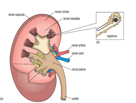
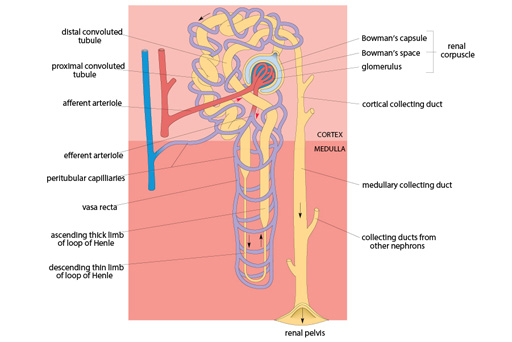
Within each kidney there are approximately one million structures called nephrons, each of which acts as an independent filter and urine-processing unit. A nephron consists of a renal corpuscle, which lies in the cortex, and a long tube which collects and processes the filtered fluid, called a renal tubule.
At the renal corpuscle, a network of very small-diameter blood capillaries, known as the glomerulus, comes into close contact with the closed end of the tubule, which is composed of a single layer of epithelial cells – the Bowman’s capsule.
In this specialised region, fluid is filtered out of the blood capillaries, across the epithelial cells and into the lumen of the tubule. The filtrate then passes along the tubule, which is convoluted (in some cases looping down into the medulla), before finally joining with the renal pelvis, where the urine is emptied into the ureters.
It is during its passage along the tubule that the contents of the filtrate are processed, and urine is formed. Most of the filtered water, glucose, amino acids, sodium and other ions are reabsorbed by the epithelial cells of the tubules. Waste substances are either not reabsorbed at all, or only partially reabsorbed.
Some molecules and ions are also secreted into the tubule by the epithelial cells and, together with waste products which remain in the filtrate, are excreted in the urine.
Cells of the kidney thus perform three main functions to produce urine:
- filtration occurs in the glomerulus/Bowman’s capsule
- reabsorption and secretion occur in the tubules.
The following diagram illustrates the blood supply to different parts of the nephron. High pressure within the glomerulus promotes the initial filtration of the blood plasma, and the network of vessels associated with the loop of Henle allows reabsorption of ions and water.