1.3 Histopathology for the diagnosis of disease
In this video, David provides some guidance for the systematic examination of slides, based on an understanding of what the normal tissue looks like.
Download this video clip.Video player: 39442_ou_futurelearn_mc1008_vid_015-540.mp4
Transcript
DAVID MALE
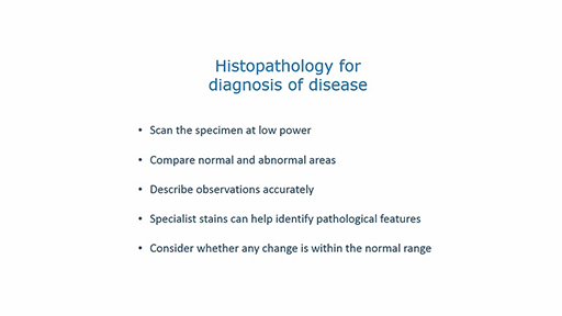
Histopathology for the diagnosis of disease. In this video, I will introduce some of the pathological specimens that you will be examining. As with any histological section, you should start by scanning the section at low power to identify any major anatomical abnormalities. The higher power objectives can be used to see changes in the individual cells or the extracellular matrix.
Sections will often have normal and abnormal areas, which can be compared. At this stage, you may not remember what the normal tissue looks like, so flip back to a normal tissue section for comparison.
When describing the appearance of a section, be absolutely clear and objective about what you see. Any interpretations can come later.
In some cases, there are pairs of sections-- one with hematoxylin and eosin stain and one with an immunohistochemical stain, which identifies specific features which may be critical for diagnosis.
When interpreting a section, consider whether any changes might be just a normal physiological change, for example, due to age or hypoplasia, and don't over interpret what you see. Remember, it takes many years to train a consultant histopathologist. Now, let's take a look at some sections.
In this case, I've loaded section number 7 from the cerebral cortex of someone who had dementia. It has been diagnosed as Alzheimer's disease. But how do we know that?
At low power, you can see the edge of the cortex and the meninges with some blood vessels. The rest of the tissue is fairly homogeneous.
I look at high power across the width of the section. I'm going to track to the left towards the edge of the cortex.
The nuclei of the neurons are small and condense, which suggests they have died, possibly by apoptosis. There is no significant infiltration with inflammatory cells, leukocytes. But as I carry on, you can see areas with vacuoles where neurons have been lost. The blood vessels appear relatively normal.
Just for comparison, I will look at a normal cortex, section 6. Although the staining here is rather different, you can see that the neurons are denser and they don't have these small condensed nuclei. They look much more normal.
Back to section 7 again. One of the hallmarks of Alzheimer's disease are plaques in the cortex containing deposits of a protein, A beta. But we can't seem them well on this H&E section. And this is where a histopathologist might ask for a specialised stain.
And section 8 is an immunohistochemical stain for A beta. You can immediately see deposits of the protein, referred to as beta amyloid, distributed throughout the cortex. If I increase the magnification, you can see that these deposits are extracellular. And they are not particularly associated with blood vessels. So this strongly indicates a diagnosis of Alzheimer's disease.
When you look at these sections, try to think about the normal anatomy and function of the tissue, as this will help in interpreting your observations.
Interactive feature not available in single page view (see it in standard view).
When viewing a section, it is important to note and report any changes objectively. Interpretation of what has caused the changes takes considerable experience. Even if you have a good idea of the underlying cause, observations and interpretation should be clearly distinguished.