3 Tumours
Cell division is normally a highly regulated process. The number of cells in any tissue is usually fairly constant, although some tissues can respond to physiological demand by an increase in cell number. Tumours disrupt this regulation.
Hyperplasia
What process occurs as mountaineers acclimatise to high altitude? The number of erythrocytes in their blood increases. But why does this happen? The fall in the level of oxygen in the air at altitude means that the capacity of the blood to carry oxygen increases, to compensate. There is a progressive increase in the number of erythrocytes over a period of weeks as the bone marrow responds by increasing production.
Other types of cell may increase in number in response to appropriate stimuli. For example, in a guitar player, the basal cells of the epidermis in the fingertips can proliferate to produce hard pads of keratin (calluses), caused by repeated contact with the strings. Cell proliferation and the consequent increase in cell numbers, seen in these two examples, is called hyperplasia. It is a normal physiological response to demand placed on a tissue.
Dysplasia
If cell division becomes poorly regulated, cells may lose some of their morphological characteristics and/or functions. The tissue becomes disordered in appearance, often with an increase in the number of immature cells, and greater variability between cells. This appearance is called dysplasia.
It should be emphasised that dysplasia does not necessarily show that the cells have become cancerous; however, it does suggest underlying changes in the cells, which may predispose them to cancer. In this sense, dysplasia may be a stage on the way to cancer development. For example, when histologists screen cervical smears, they are particularly looking for changes in the normal morphology of the cells which indicate pre-cancerous changes.
Neoplasia
Neoplasia is the term used to describe the development of tumours or cancerous tissue.
The development of a tumour requires a series of changes in the biology of the cell, with progressive loss of the controls that limit cell division. Even a cell that is undergoing uncontrolled proliferation will not necessarily be malignant. Malignancy typically arises when the dividing cells invade the normal tissue and move away from their site of origin. Because of the great variety of different tumours, it is impossible to generalise. Nevertheless, it is very important for a pathologist to be able to distinguish between a benign tumour and a malignant cancer, since the treatment required will usually be radically different. Consequently, pathologists often grade tumours according to how malignant/invasive they are.
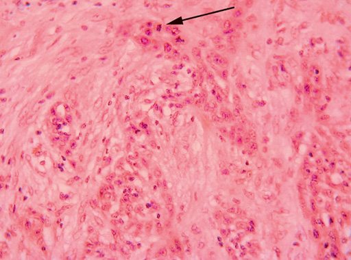
Histologists can get some impression of the rate of cell division within a tissue, according to the number of mitotic figures – the number of cells showing the characteristic pattern of separating chromosomes, seen as the cell divides. Invasion of tumour cells within the tissue can be estimated, by observing where the cells are in relation to their normal position and in relation to other cells in that tissue, and this forms an important element in the pathological report on a tumour.