

3.1 Absorption into the blood
Once the food has been digested, the next step is for the products – sugars, amino acids, fatty acids and monoacylglycerols – to be absorbed (see Figure 9).
The inside of the small intestine is covered in minute, finger-like projections, called villi (Figure 9). This makes the lining look and feel – but not smell – like velvet. These villi give the lining of the small intestine a huge surface area, calculated to be about 30 m2, over which the digested food can be absorbed.
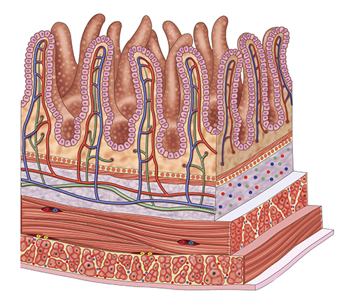
Capillaries supply blood to each villus. The blood brings oxygen to keep the cells of the intestine alive and absorb and take away the sugars and amino acids. These tiny capillaries join together to form a vein called the hepatic portal vein, which takes all of the blood to the liver. The fatty acids and monoacylglycerols are absorbed, not into the blood directly, but into blunt-ended tubes in the villi called lymphatic vessels. These all join together and the fluid in them empties into the blood system close to the heart.