1.4 Immunohistochemistry
In the last 30 years, staining methods using antibodies have been increasingly developed. When applied to staining for the light microscope, these techniques are collectively referred to as immunocytochemistry (ICC) for the staining of cells or immunohistochemistry (IHC) for the staining of tissues.
Antibodies are proteins synthesised by one of the cell types of the immune system, but their use for staining other cells has revolutionised histology, as they allow the identification and localisation of individual molecules. Antibodies specifically bind and detect individual proteins. They can therefore be selected to identify the presence or location of a single protein that identifies a single cell type, or is diagnostically discriminating.
A molecule that binds to, and is recognised by, an antibody is called an antigen, and, in the context of histology, such antigens are often referred to as markers, since they act as ways of recognising a particular cell.
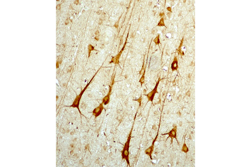
Staining with antibodies is particularly valuable for distinguishing between different cell types in a tissue. For example, different classes of lymphocyte appear virtually identical in size and shape, but they can be distinguished through staining according to their surface markers. Similarly, in Figure 7 some nerve cells (neurons) are clearly visible among other cells in the cortex of the brain as a result of this type of staining technique.