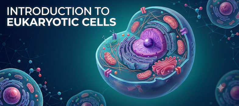
B. Structure of the Eukaryotic Cell
1. The Plasma Membrane
The plasma membrane is an essential and ubiquitous structure, as all cells must possess one. It performs both crucial structural and dynamic roles. Structurally, it acts as the definitive boundary that segregates the cellular material from the environment, helping to maintain cellular integrity. This integrity is frequently aided by other associated structures, such as rigid cell walls or extensive internal and external protein matrices. Dynamically, the membrane facilitates several key processes, including regulated transport of substances, communication with the cell's environment, and cellular motility.
Composition and Structure
The architecture of the plasma membrane is best understood as a fluid matrix composed mainly of lipids and proteins.
Lipids:
Phospholipids and other membrane lipids form the fundamental bilayer structure and are often the most abundant molecular component in many membranes. These are amphipathic molecules, meaning they possess a hydrophobic side (the tail) and a hydrophilic side (the head). This intrinsic duality dictates the bilayer arrangement, which features a non-polar hydrophobic core that serves as an effective barrier to the diffusion of polar molecules. The hydrophilic sides are exposed to the surrounding environment, perfectly suited to separate the internal and external aqueous solutions.
The molecular structure includes several classes of lipids:
- Glycerophospholipids (aka Phosphoglycerides): These are characterized by two fatty acids, each with a long hydrophobic tail and a hydrophilic carboxylic acid, a hydrophilic glycerol-phosphate backbone, and a variable hydrophilic head group (e.g., amino alcohols, amino acids, polyalcohols, or sugars). A common example is Phosphatidylcholine (PC), which features choline as its head group.
- Sphingolipids: These are based on the fatty amino alcohol sphingosine and also contain a phosphate, one fatty acid, and a head group. Sphyngomyelin, with choline as its head group, is a major example.
- Cholesterol Family (Sterols): These are molecules with a small hydrophilic head and a bulky hydrophobic side containing multiple rings. Their function is to enhance the physical stability and increase the rigidity of the membrane. Cholesterol is the key example in animal membranes.
Proteins:
Membrane proteins confer specificity and dynamic capabilities to the lipid bilayer:
- Integral Proteins: These are embedded directly within the membrane, interacting with the hydrophobic core. Those that fully cross the membrane are termed transmembrane proteins. Their membrane-spanning domain typically consists of an alpha helix composed of predominantly hydrophobic amino acids. A well-studied example is the Glycophorin A dimer.
- Peripheral Proteins: These are attached to the surface of the membrane, interacting primarily with the hydrophilic sides (the lipid heads or other integral proteins). They are essential for conferring many of the membrane's dynamic functions, such as transport, communication, and metabolism.
Fluid Mosaic Model and Asymmetry
The accepted structural paradigm is the Fluid Mosaic Model, which describes the membrane as a mosaic of proteins suspended within a dynamic lipid bilayer. The membrane is fluid, allowing for the lateral movement of both proteins and lipids. This fluidity is subject to control by temperature and lipid composition.
Furthermore, biological membranes exhibit significant asymmetry, meaning they are composed of two different layers, or leaflets (the exoplasmic and cytoplasmic faces), with distinct compositions.
- Lipid Asymmetry: For instance, Phosphatidylserine (PS) is preferentially located in the cytoplasmic leaflet, while glycolipids are typically found in the exoplasmic leaflet.
- Protein Asymmetry: Proteins exhibit a very specific orientation relative to the cell's interior and exterior. For example, Glycophorin A consistently has its glycosylated N-terminal domain facing outside the cell and its C-terminal domain facing inside.
2. The Nucleus and Cytoplasm of Eukaryotic Cells - Overview
Organelles
Eukaryotic cells are defined by an extensive system of internal biomembranes that form numerous membrane-bound organelles. This feature facilitates intracellular compartmentalization, which in turn enables specialized functionality due to the unique biochemical composition of each compartment.
Key membrane-bound organelles include:
- Nucleus: Houses the cellular Genome.
- Lysosomes: Centers for degradation via acid hydrolases.
- Vacuoles: Primarily used for storage (especially large in plants).
- Peroxisomes: Involved in degradation via oxidases and detoxification.
- Mitochondria: The sites of aerobic respiration and primary energy metabolism.
- Chloroplasts: Sites of photosynthesis (in plants and algae).
- Endoplasmic Reticulum (ER): Responsible for the biosynthesis of membrane proteins and lipids, secretion, and detoxification.
- Golgi Apparatus: The key center for trafficking, distribution, and secretion of proteins and lipids.
Cytosol
The cytosol is the aqueous, dense fluid that fills the cell, sometimes visualized as a "soup" of biomolecules. It contains various small biomolecules and macromolecules, serving functions related to storage, metabolism, structure, and motility.
3. The Nucleus
Structure and Composition
The nucleus is typically the largest organelle in animal cells. It is bounded by a double membrane known as the nuclear envelope. The inner nuclear membrane faces the nucleus content, while the outer nuclear membrane is continuous with the ER. Both membranes are crossed by nuclear pores, lined by large protein complexes (NPCs) that actively regulate transport between the nucleoplasm and the cytoplasm.
The content of the nucleus, the nucleoplasm, contains the genetic material packaged as Chromatin, which is DNA complexed with proteins. Chromatin is classified based on density: Euchromatin is less densely packed and appears lighter, while Heterochromatin is more densely packed and appears darker. Specialized regions called the Nucleolus or nucleoli are the primary centers for rRNA synthesis.
Function (Activity)
The nucleus is always an active organelle. Replication of the DNA occurs here, though this is restricted to cells preparing to divide. Transcription (the synthesis of RNA from DNA) is a critical activity that occurs in all living cells.
4. Lysosomes
Structure and Composition
Lysosomes exist as two main types: Primary lysosomes are small, round, and regular vesicles, while Secondary lysosomes are larger and more irregular. They are bounded by a single lysosomal membrane that is equipped with an ion pump transporter (H+). This pump, assisted by a Cl- channel, actively maintains a highly acidic pH (<5) within the lumen. The lumen contains a powerful cocktail of acid hydrolases capable of degrading diverse macromolecules, including nucleic acids, polysaccharides, proteins, and lipids.
Function
Lysosomes are the cell's essential machinery for intracellular digestion and recycling. Their functions include:
- Endocytosis: Degradation of materials delivered in small endosome vesicles.
- Phagocytosis: Degradation of large particles ingested in phagosomes.
- Autophagocytosis: The essential process of recycling the cell's own organelles and damaged components.
5. Vacuoles
Structure and Composition
Vacuoles are single-membrane sacs that are highly variable in size. They are particularly prominent in plant cells, where they often form a very large central organelle. The boundary membrane is the vacuolar membrane or tonoplast, which contains various transporters. The lumen holds a diverse mixture of materials, including water, ions, small organic compounds, and macromolecules.
Function
Vacuoles are extremely important in plants. They function in the storage of various compounds, including waste products. Crucially, they hold water at high pressure, which is vital for providing turgor (structural support) and is necessary for growth through the lengthening of cells.
In animal cells, vacuoles are typically much less important, often being small and transient. A unique exception is the pulsatile vacuole found in protozoans, which functions in water pumping to control osmotic pressure, thus allowing the organism to thrive in freshwater environments.
6. Peroxisomes
Structure and Composition
Peroxisomes are small membranous sacs or vesicles (one of the last organelles to be discovered, circa 1954/1967). They are enclosed by a single membrane that includes various transporters, notably one specific for very long chain fatty acids. The lumen contains specialized enzymes, primarily Oxidases and Catalase.
Function
The core function of peroxisomes is the oxidation of compounds using molecular oxygen. This includes the breakdown of very long chain fatty acids, mainly to generate precursors rather than to produce ATP (the energy is released as heat). Defects in this process are linked to conditions such as Adrenoleukodystrophy (ALD). Peroxisomes are also crucial for detoxification, particularly the degradation of toxic compounds in the liver. A highly toxic byproduct of their oxidative reactions, hydrogen peroxide (H2O2), is generated, and its prompt removal is ensured by the enzyme Catalase to prevent cellular damage.
7. Mitochondria
Structure and Composition
Mitochondria are large organelles characterized by a complex structure bounded by two membranes. The outer mitochondrial membrane is relatively permeable to small molecules, typically composed of 50% lipid and 50% protein. The inner mitochondrial membrane is extensively invaginated, forming folds called cristae, which dramatically increase its surface area. This inner membrane is highly protein-rich (approximately 70%), housing the components of the Electron Transport Chain and ATP synthase. The innermost compartment, the matrix, contains enzymes necessary for metabolism (e.g., Pyruvate Dehydrogenase, Beta-oxidation pathway enzymes, and Krebs cycle enzymes), as well as their own Ribosomes (70S) and a molecule of circular DNA, structural evidence consistent with the Endosymbiont theory.
Function
Mitochondria function as the major ATP synthesis center of the cell. They are responsible for the catabolism of sugars and fatty acids through aerobic respiration metabolism, which includes the Krebs cycle and Oxidative Phosphorylation (mediated by the Electron Transport Chain and ATP synthase).
8. Chloroplasts
Structure and Composition
Chloroplasts are very large, complex organelles found only in plant and algae cells. They are bounded by three membranes: an outer membrane, an inner membrane, and specialized internal membranes. The internal membranes, known as thylakoid membranes, form sacs that often stack into structures called grana. These membranes are protein-rich, containing the Photosystems, the Electron Transport Chain, and ATP Synthase. The fluid-filled interior, the stroma, contains enzymes such as those for the Calvin cycle, Ribosomes (70S), and circular DNA, also supporting the Endosymbiont theory.
Function
The primary function of chloroplasts is Photosynthesis. Light energy is captured at the thylakoid membranes to generate high-energy molecules, specifically NADPH and ATP. This energy is subsequently used in the stroma to convert carbon dioxide (CO2) into sugar precursors.
9. The Endoplasmic Reticulum (ER)
Structure and Composition
The Endoplasmic Reticulum (ER) is a vast, interconnected network of sacs. It is bounded by a single membrane and represents the largest total amount of intracellular membrane in eukaryotic cells. It is structurally divided into two types: the Smooth ER (which is lipid-rich) and the Rough ER (which is protein-rich and visibly contains ribosomes attached to its surface under electron microscopy). The internal space, the lumen, is rich in enzymes capable of modifying both lipids and proteins.
Function
The ER performs multiple functions and serves as the start of the secretory pathway. Key functional divisions include:
- Smooth ER: Primarily responsible for the biosynthesis of membrane lipids and the detoxification of hydrophobic molecules.
- Rough ER: Primarily responsible for the biosynthesis of membrane proteins and the synthesis of proteins that follow the secretory pathway, as well as participating in their initial protein maturation and modification.
10. The Golgi (Apparatus)
Structure and Composition
The Golgi apparatus is a structure composed of a series of flattened sacs (cisternae) and associated small vesicles, all enclosed by a single membrane. It is functionally divided into three regions, each containing specific enzymes: the Cis Golgi, the Medial Golgi, and the Trans Golgi. The internal lumen is rich in enzymes that can modify lipids or proteins.
Function
The Golgi acts as the cell's processing and distribution center, functioning as a critical part of the secretory pathway. It receives small vesicles from the ER. Proteins and lipids are then sequentially modified as they traffic through the complex (Cis > Medial > Trans Golgi). The Trans Golgi is responsible for sorting and sending small vesicles to their final cellular targets, which include lysosomes, the plasma membrane, or for secretion to the extracellular environment through secretory vesicles.
11. The Cytosol
Structure and Composition
The cytosol is a dense, fluid environment, rich in proteins and RNAs, structurally akin to a concentrated "soup". Approximately 70% of the cytosol is liquid, primarily H2O, containing dissolved solutes like sugars, amino acids, and nucleotides. The remaining 30% consists of suspended or dispersed solid materials, mainly proteins and protein complexes. These include numerous enzymes and enzyme complexes, Ribosomes, and an extensive intracellular network of protein fibers known as the Cytoskeletal fibers. This network is composed of three types of protein fibers: the very thin microfilaments (7 nm), the intermediate filaments (10 nm), and the thicker microtubules (25 nm). The cytosol also contains larger, insoluble structures referred to as inclusion bodies (the "chunks in a soup"), such as Glycogen particles (prevalent in liver and muscle cells) and large Triglyceride droplets (abundant in fat cells, or adipocytes).
Function
The cytosol serves multiple critical roles, including Storage, acting as the site for central pathways of cellular Metabolism, hosting Protein synthesis, and providing both Structure and motility through the action of the cytoskeleton.