Acknowledgements
The course image and banner were generated with Google Gemini.
Prompts:
Course Image
Subject: A high-resolution, professional educational infographic titled "THE ANATOMY AND PHYSIOLOGY OF EUKARYOTIC CELLS." The layout should be clean, organized, and scientifically accurate, divided into three distinct panels.
Panel 1: The Fluid Mosaic Model (Top Left)
-
Visual: A detailed 3D cross-section of a plasma membrane.
-
Components: Show a phospholipid bilayer with hydrophilic heads and hydrophobic tails. Include integral membrane proteins (e.g., Glycophorin A as a homodimer and a single transmembrane monomer).
-
Annotation: Display "Lipid asymmetry: Phosphatidylserine" and labels for "Hydrophilic heads," "Hydrophobic core," and "Extracellular Domain (glycosylated)."
-
Style: Clean, textbook-style scientific illustration.
Panel 2: The Secretory Pathway (Bottom Left)
-
Visual: A 3D illustration of the nucleus, endoplasmic reticulum (RER), and Golgi apparatus.
-
Nucleus: Include clearly visible nuclear pores on the surface, with euchromatin and heterochromatin visible inside. Important: No labels or arrows should be pointing inside the nucleus or toward the nucleolus. Only clear, distinct labels for "Nuclear pores," "Euchromatin," and "Heterochromatin."
-
Pathway: Show the flow from RER to Golgi via transition vesicles. The cis-Golgi must be placed closest to the RER. The stack should be labeled "Golgi apparatus" with sub-labels for "medial" and "trans" cisternae. Indicate "Exocytosis" occurring at the trans-Golgi face.
Panel 3: Integration of Structure and Function (Right Side)
-
Visual: A vertical arrangement of three distinct organelle icons layered over a soft-focus electron micrograph background.
-
Top Icon: Mitochondrion with an energy/lightbulb icon, labeled "Energy/Mitochondrion."
-
Middle Icon: Lysosome (a green circular organelle) with a recycling symbol positioned immediately above the text label "Degradation/Recycling."
-
Bottom Icon: Chloroplast with a sun icon, labeled "Chloroplast: Photosynthesis."
-
Constraint: There should be no connecting lines or arrows between these organelles and the electron micrograph background.
Overall Style and Formatting:
-
Aesthetic: Clear, sans-serif typography, high contrast, professional academic color palette (soft blues, greys, and distinct accent colors for cellular structures).
-
Composition: Balanced white space, clear section headers, and logically grouped annotations.
-
Technical Requirement: Ensure all anatomical structures are rendered with scientific accuracy consistent with collegiate-level biology textbooks.
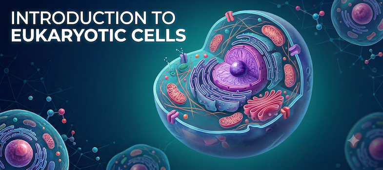
Course Banner
A crisp, high-resolution course header banner (785x350 aspect ratio, widescreen layout) titled "INTRODUCTION TO EUKARYOTIC CELLS" displayed prominently on the left side in modern, bold, clean white sans-serif typography.
The central focus of the image is a highly detailed, 3D cross-section illustration of a eukaryotic cell floating on a deep teal and navy gradient background subtle grid network. The cell is cut open to clearly display its internal organelle architecture with vibrant, distinct coloring and soft luminescence:
-
Nucleus: A central purple sphere with an exposed inner nucleolus and rich chromatin textures.
-
Endoplasmic Reticulum (ER): Folded, layered blue membranous sheets surrounding the nucleus.
-
Golgi Apparatus: Curved pink and magenta flattened sacs positioned near the edge.
-
Mitochondria: Bright red, oval-shaped powerhouses showing inner cristae folds.
-
Cytoskeleton & Plasma Membrane: A clear, semi-translucent outer phospholipid bilayer shell supported by fine internal cytoskeletal filaments.
Soft molecular structures, glowing blue/magenta micro-particles, and out-of-focus background cells add depth to the composition. Clean, professional educational graphic design, cinematic lighting, vibrant scientific visualization, macro detail. No extra text, no labels, no pointers, no subheadings.