Figures: Structure of the Eukaryotic Cell
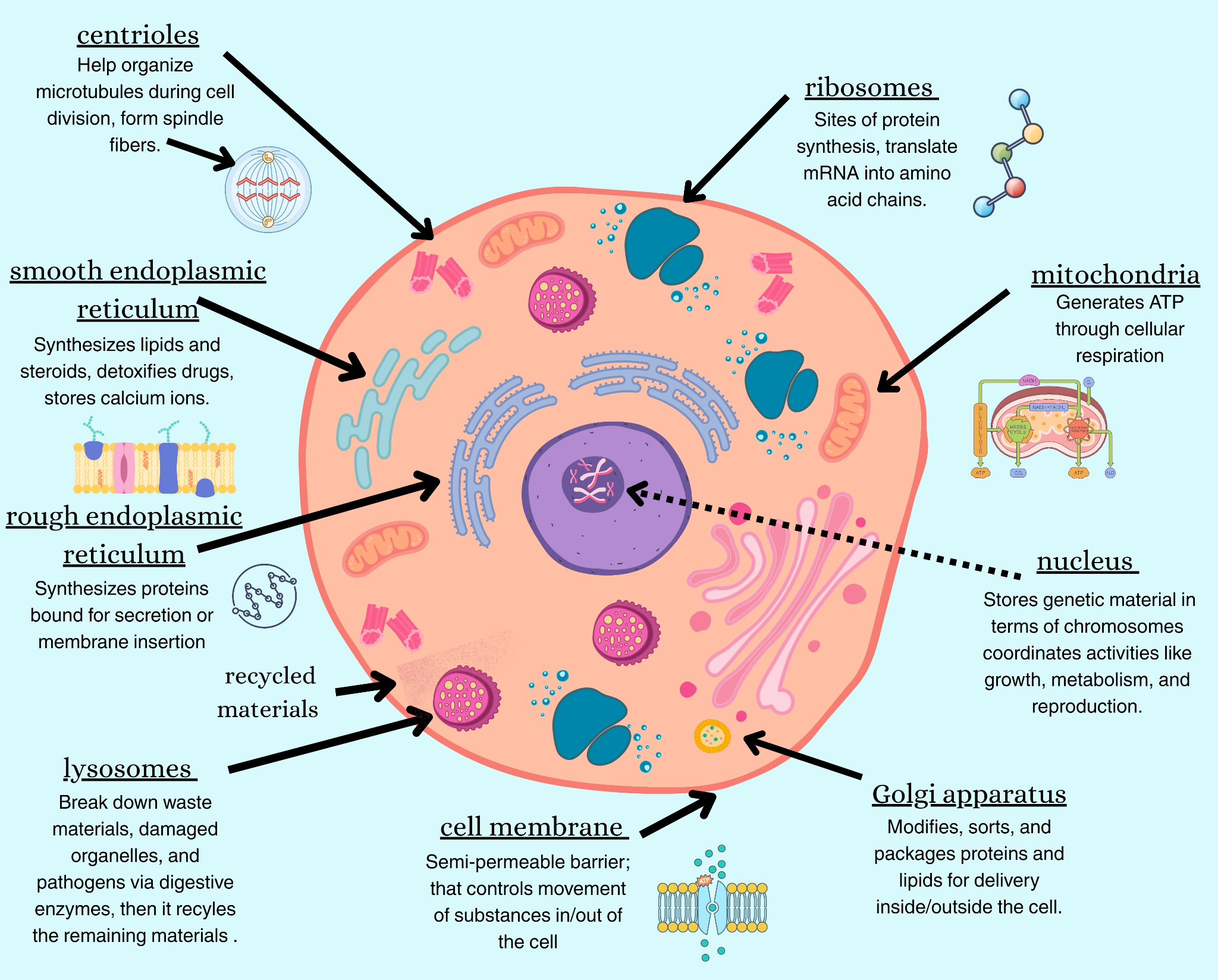
Figure 1. Schematic diagram illustrating the key components of a generalized eukaryotic cell. Major organelles, including the nucleus, mitochondria, endoplasmic reticulum, and Golgi complex, are labeled to demonstrate the characteristic compartmentalization of eukaryotic cell structure.
Source: Eukaryotic cell diagram | License: CC BY-SA 4.0

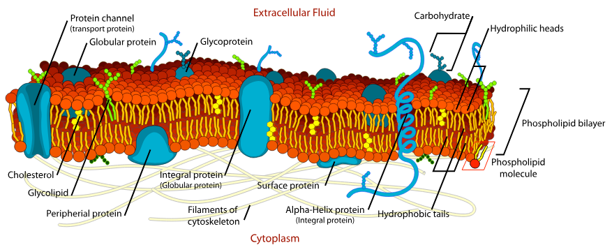
Figure 2. A detailed, annotated diagram illustrates the fluid mosaic model of the eukaryotic cell membrane, highlighting the phospholipid bilayer, various integral and peripheral proteins, cholesterol, and associated glycoproteins and glycolipids. The image displays the asymmetric distribution of these components, which facilitates specific membrane functions like transport and cell recognition.
Source: Cell membrane diagram | License: Public domain

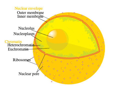
Figure 3. Schematic diagram illustrating the detailed ultrastructure of a human cell nucleus. Key components depicted include the double-membraned nuclear envelope, nuclear pore complex, chromatin, nucleoplasm, and the central nucleolus.
Source: Diagram human cell nucleus | License: Public domain
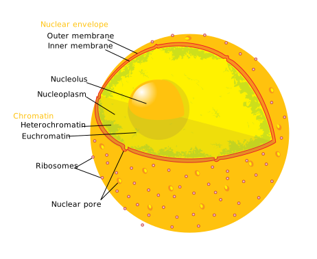
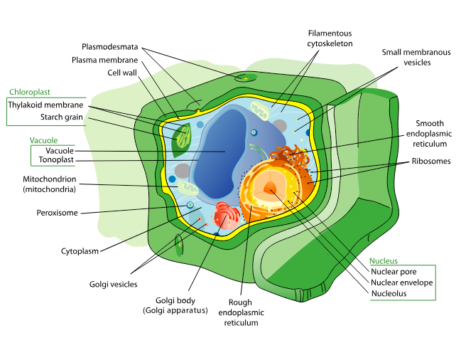
Figure 4. A detailed, labeled diagram illustrating the major organelles and structures of a typical eukaryotic plant cell, including the cell wall, cell membrane, nucleus, vacuole, chloroplasts, and mitochondria. This figure provides a comprehensive overview of the spatial organization and key components of plant cellular anatomy.
Source: Plant cell structure | License: Public domain

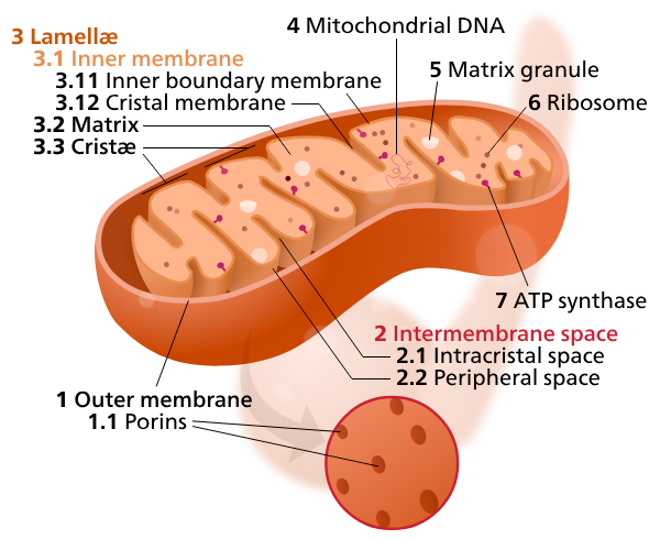
Figure 5. Simplified schematic diagram illustrating the double-membrane structure of a typical mitochondrion. Key structural components shown include the outer membrane, the highly folded inner membrane (cristae), the intermembrane space, and the internal matrix.
Source: Mitochondrion | License: CC0
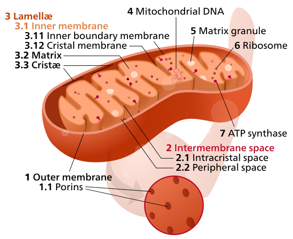
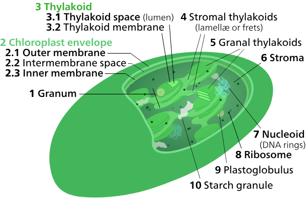
Figure 6. Diagrammatic representation of a chloroplast, the organelle responsible for photosynthesis in eukaryotic cells. Key features include the double membrane envelope, the fluid-filled stroma, and the internal stacked thylakoid membranes (grana).
Source: Chloroplast | License: CC BY 3.0
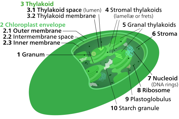
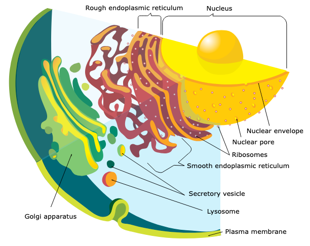
Figure 7. This diagram illustrates the components and functional connectivity of the eukaryotic endomembrane system, encompassing the endoplasmic reticulum, Golgi apparatus, lysosomes, and various transport vesicles. The figure highlights the sequential flow of synthesized macromolecules from the endoplasmic reticulum through the Golgi for modification and subsequent delivery to their final cellular destinations.
Source: Endomembrane system diagram | License: Public domain

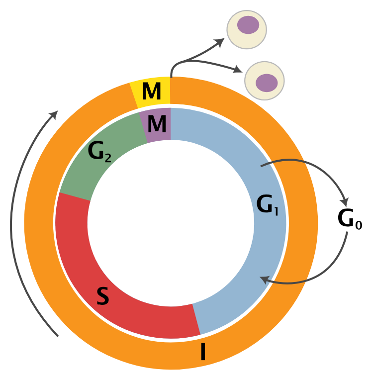
Figure 8. A diagram illustrating the eukaryotic cell cycle, detailing the progression through interphase (G1, S, and G2 phases) and the mitotic (M) phase. This representation highlights the sequential nature of growth, DNA synthesis (S phase), and cell division, including the option for cells to enter the quiescent G0 state from G1.
Source: Cell Cycle | License: CC BY-SA 3.0
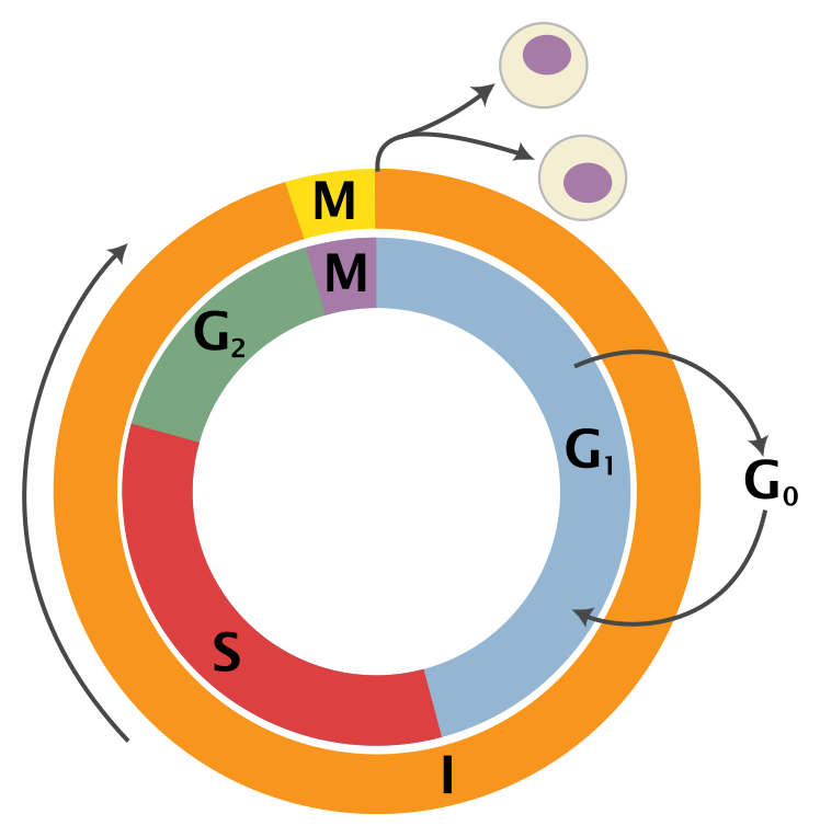
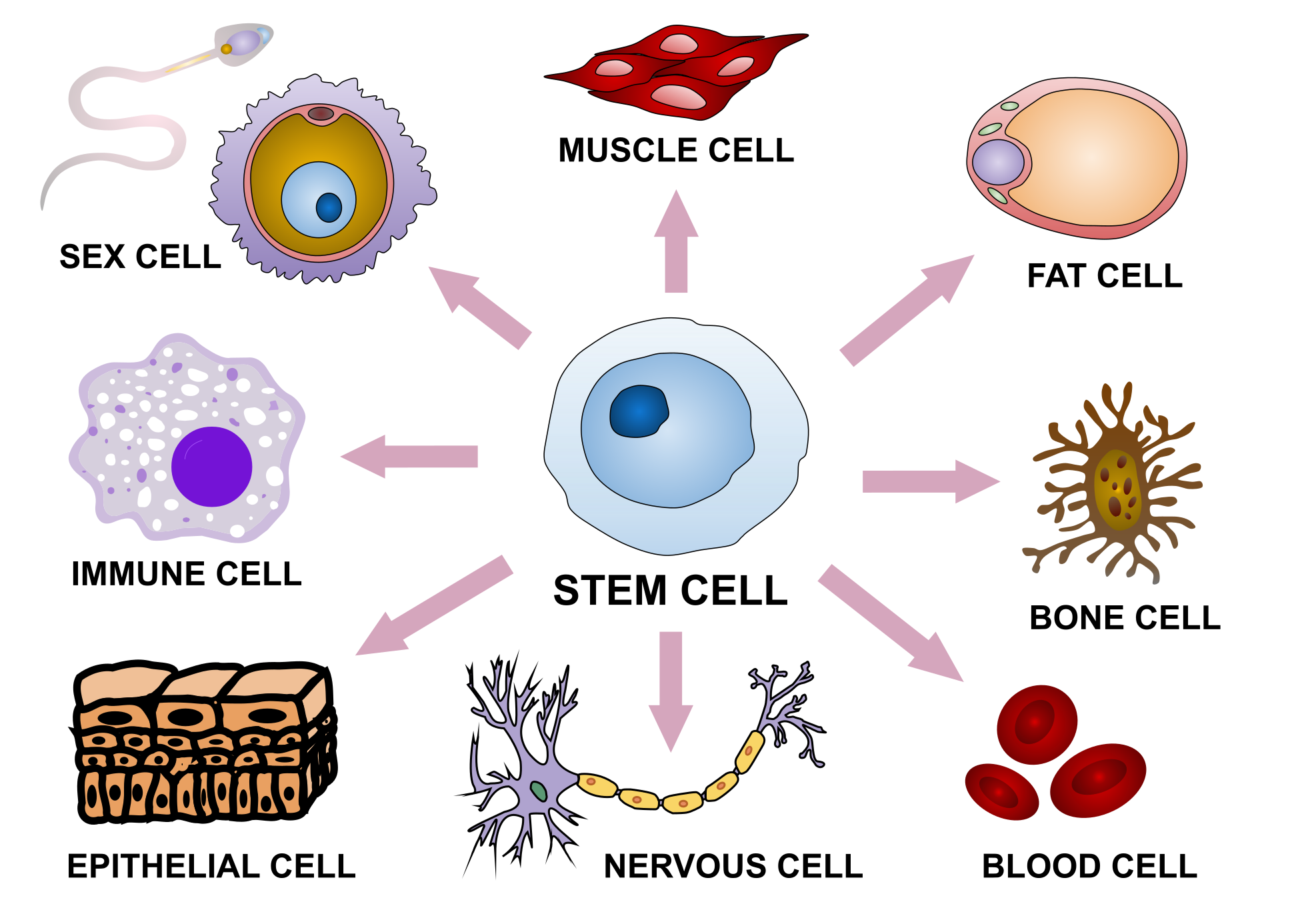
Figure 9. The diagram illustrates the model of cell differentiation from highly versatile totipotent stem cells to the ultimate formation of specialized cell types.
Source: Cell Differentiation | License: CC BY-SA 4.0

Click here to take the course quiz