

4 Milk production (lactation)
In this section, you are presented with a fairly complex diagram, Figure 1. The first thing to do when you come to any diagram is to read the caption (i.e. the title), which explains what it shows. Then look at the diagram itself, taking particular note of the scale, if there is one. You may find that the accompanying text 'talks you through' the diagram step by step, as it does here. Visualising three-dimensional objects from a two-dimensional diagram is a difficult skill and you should not expect to master it straight away, so don't be concerned if you don't immediately understand all aspects of Figure 1.
I'll now say more about one of the defining features of mammals - milk production. This feature isn't talked about in any detail in LoM but it's such a remarkable and unique mammalian process that it warrants attention here. The production of milk (technically called lactation) makes sense only if we look inside mammals to find out how this life-sustaining substance is produced.
Milk is a very rich form of food. You've probably already heard about some of the major constituents of milk - proteins, fats and carbohydrates. These large molecules have to be built up (synthesised) from the simpler chemicals that the mother obtains from her diet or from her body reserves. By looking at the structure of a typical mammary gland we'll see how this biological 'production line' is put together.
The term 'gland' is used for specialised structures that produce (or more technically secrete) one or more chemical products, and many glands have the type of structure that Figure 1 shows. (Glands are usually made up of different types of cells - a group of cells that have similar structure and function is often called a tissue.)
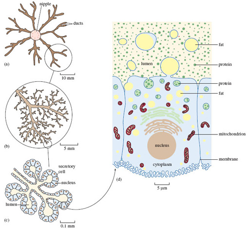
Figure 1a shows that each mammary gland consists of a central teat or nipple, into which feed a number of channels (or ducts) that convey and temporarily store the milk, following its production by the great mass of cells that make up the bulk of the gland. We get a better sense of the fine-detailed structure of the mammary gland by magnifying just one part of what's shown in (a). You'll appreciate that all the ducts shown have much the same structure, but focusing on just one makes things clearer. Figure 1b shows one representative part, around the blind ends of the finely branched ducts. At this magnification, the individual secretory cells aren't visible; but you can make them out in Figure 1c, a higher-powered view of just one small part of (b). In (c) you can see five such small groups, shown in a schematic way in cross-section - in other words, it's a 'tidied up' view of an imaginary slice though the gland. If you imagine these structures in three dimensions, (c) would resemble a 'bunch of grapes', the stalks representing the ducts and each 'grape' a group of secretory cells surrounding a central space (or lumen), which would contain milky fluid.
The structure of each individual cell in (c) is much the same and resembles that of many other cells in the mammalian body. Each cell is roughly rectangular in cross-section, with a thin outer membrane. Towards the base of the cell is a small rounded nucleus, which contains most of the genetic material (the DNA), which has a key role in directing the workings of the cell.
Figure 1d is a hugely magnified view of just one of the secretory cells shown in (c) - a single sample of the many millions of cells that comprise the mammary gland. Magnification on this scale requires the use of an electron microscope, as opposed to the less powerful optical light microscope used for (a)-(c) that you might be more familiar with. You'll recognise the cell's roughly rectangular shape and the rounded nucleus; you can also see the cell membrane.
The identity and function of all the many different cellular components in (d) needn't concern us in detail. There are many flattened, highly folded membranes, resembling stacked piles of plates. A few of these are shown in (d), just above the nucleus. (You will notice that there are two types of internal membrane stacks, denoted here in different colours.) The sausage-shaped structures are called mitochondria (singular: mitochondrion) - often termed the powerhouses of the cell, because they deliver the energy that fuels the complex synthetic processes that I'll talk more of in a moment. Towards the upper part of the cell, closest to the lumen, there are a number of fluid-filled droplets (called vesicles), containing what look like small granules. In reality, they are mostly different types of protein - just one of a range of large molecules (i.e. macromolecules) that make up the chemical constituents of all living material. In fact, I've drawn attention to proteins in Figure 1d by showing them as granules; though even at this level of magnification, proteins would not be discernible, since they are soluble in the fluid that contains them. Milk contains many proteins and most of them are assembled into these complex structures from much simpler chemical building blocks (called amino acids) within the secretory cells like the one shown in (d). The proteins in milk are vital to the growth and wellbeing of the suckling and some (collectively called antibodies) help the youngster withstand infection.
Fat is another key constituent of milk. You'll know that the fat content of milk can vary over time [p. 32], and the variation in milk composition between species is even greater. For example, rhinoceros milk contains hardly any fat, while seal milk is almost 50% fat. In those species where milk production has been most thoroughly investigated, secretory cells in the mammary glands take up fats from the bloodstream. (They also synthesise fats from other nutrient molecules carried to them in the blood, such as sugars.) The basic constituents of milk fats are then assembled together, most of them in parallel stacks of folded membranes of the type shown in Figure 1d. Fat accumulates within the droplets evident in (d), in the process of migrating to the upper part of the cell. The fat droplet, itself wrapped in a membrane, merges with the cell membrane and is 'budded-off' - fat droplet plus its enveloping membrane - into the lumen. From (d) you'll appreciate how each such structure resembles a decorative ring, with a fragment (of cell) often protruding from the central sphere - hence the name 'signet'.
Figure 1d conveys a sense of great cellular activity - all these synthetic processes require building blocks and the input of energy. These demands explain why lactation is a very considerable 'investment' by the mother in the wellbeing of her offspring. For animals on an unreliable or low-energy diet - the koala is a good example of the latter - synthesis of the constituents of milk can be a precarious operation. Given that the energy (fat) content of koalas' milk is comparatively low, it's not surprising that the period of lactation in koalas is unusually long, even by marsupial standards. At the age of five months, young koalas start to ingest eucalyptus leaves that have been already partly digested by the mother.
Lactation in marsupials has a particular importance; for example, the newborn red kangaroo (like the koala newborn mentioned in Section 3) weighs less than a gram - or, in the more familiar language of the TV commentary, 'less than a lump of sugar'. On complete emergence from the pouch, some eight months later, it weighs about four to five kilograms. It may then often double in weight before becoming fully independent of the mother's milk (i.e. becoming weaned), which happens between four and eight months after leaving the pouch. The composition and flow of milk are tightly controlled; the synthesis of particular nutrients can become switched on as particular genes are activated. (Such genes comprise particular sections of DNA within the nucleus of secretory cells, so minute that not even the magnification of Figure 1d can reveal them.) Towards the end of its time in the pouch, when the rate of the youngster's growth is very high and energy demands greatest, the fat content of the milk in many species reaches a maximum.
At a stage in development where hair growth is important, the youngster's production of the protein keratin has to be stepped up - keratin is the key ingredient of hair and hair-like structures. At such a time, the kangaroo's milk contains large amounts of the particular types of amino acid that are essential for the synthesis of keratin.
Question 4
Kangaroos and wallabies generally have four teats within the pouch, though there is only one newborn at a time. Why does the possession of more than one teat make sense?
Answer
Because such species have a unique and sophisticated system that allows the production of more than one type of milk, so satisfying the differing demands of youngsters at different stages of development [p. 32]. The mechanisms in the mother's body are not well understood but having separate 'plumbing' to each teat means each can act partially independently.
There's no doubt that the evolution of lactation was crucial to the success of mammals. Of course, milk production is just one component of a 'package' of parental care measures shown by many mammals, so it's perhaps risky to single out a particular feature, but it's one that raises particular interest.
Question 5
Can you think of some advantages of the lactation habit in mammals?
Answer
Lactation provides nutritional care - the sucklings don't have to find their own food. In that sense, it helps separate the infant from the vagaries of the environment. Given that feeding is relatively effortless, the youngster's energy can be largely channelled into growth. It also seems that suckling is an excellent way of transferring immunity from mother to offspring. You may know that the early milk of many mammals, humans included, confers a special advantage - it's especially rich in antibodies. The same is true in marsupials such as kangaroos. Clearly, the young are also protected in a variety of ways; often the protection is physical - remember the suckling marsupials from the TV programme.