

5 Milk production (lactation)
One of the defining features of mammals is milk production. It is a remarkable and unique mammalian process technically called lactation, which only makes sense if we look inside mammals to find out how this life-sustaining substance is produced.
Milk is a very rich form of food. You’ve probably heard about some of the major constituents of milk – proteins, fats and carbohydrates. These large molecules have to be built up (synthesised) from the simpler chemicals that the mother obtains from her diet or from her body reserves. By looking at the structure of a typical mammary gland, you can see how this biological ‘production line’ is put together.
The term ‘gland’ is used for specialised structures that produce (or more technically secrete) one or more chemical products, and many glands have the type of structure that Figure 17 shows. (Glands are usually made up of different types of cells – a group of cells that have similar structure and function is often called a tissue.)
Examining diagrams
The first thing to do when you come to any diagram is to read the caption, which explains what it shows. Then look at the diagram itself, taking particular note of the scale, if there is one. You may find that the accompanying text ‘talks you through’ the diagram step by step, as it does here. Visualising three-dimensional objects from a two-dimensional diagram is a difficult skill and you should not expect to master it straight away, so don’t be concerned if you don’t immediately understand all aspects of Figure 17.
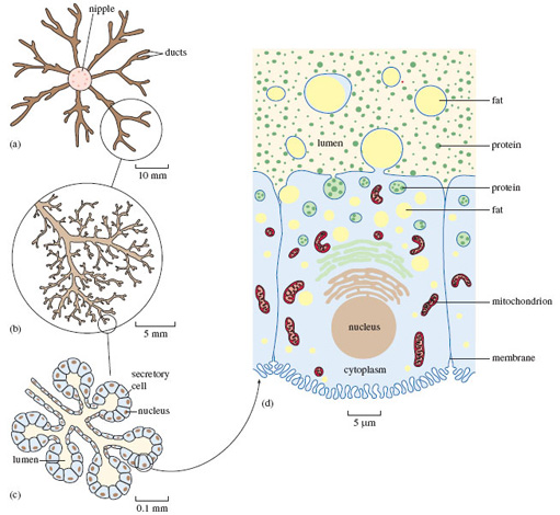
Figure 17 shows different levels of detail at increasing magnification. The image labelled (a) shows that each mammary gland consists of a central teat or nipple, into which feed a number of channels (or ducts) that convey and temporarily store the milk, following its production by the great mass of cells that make up the bulk of the gland. Magnifying just one part of what’s shown in (a) gives you a better sense of the fine-detailed structure of the mammary gland. All of the ducts shown have much the same structure, but focusing on just one makes things clearer. (b) shows one representative part, around the blind ends of the finely branched ducts. At this magnification, the individual secretory cells aren’t visible; but you can make them out in (c), a higher-powered view of just one small part of (b). In (c) you can see these small groups of cells, shown in a schematic way in cross-section. In other words, it’s a ‘tidied up’ two-dimensional view of an imaginary slice though the gland. If you imagine these structures in three dimensions, (c) would resemble a bunch of grapes, with the stalks representing the ducts and each ‘grape’ a group of secretory cells surrounding a central space (or lumen), which would contain milky fluid.
The structure of each individual cell in (c) is much the same, resembling that of many other cells in the mammalian body. Each cell is roughly rectangular in cross-section, with a thin outer membrane. Towards the base of the cell is a small rounded nucleus, which contains most of the genetic material (the DNA), which has a key role in directing the workings of the cell.
Figure 17 (d) is a hugely magnified view of just one of the secretory cells shown in (c) – a single sample of the many millions of cells that comprise the mammary gland. Magnification on this scale requires the use of an electron microscope, as opposed to the less powerful optical light microscope used for (a)-(c). Here you can see the cell’s roughly rectangular shape and the rounded nucleus, as well as the cell membrane.
The identity and function of all the many different cellular components in (d) needn’t be covered in detail. There are many flattened, highly folded membranes, resembling stacked piles of plates. Two types of these are denoted here in different colours, just above the nucleus. The sausage-shaped structures are called mitochondria – these are often termed the powerhouses of the cell, because they deliver the energy that fuels the complex synthetic processes which will be explained further in a moment. Towards the upper part of the cell, closest to the lumen, there are a number of fluid-filled droplets (called vesicles), containing what look like small granules. They are mostly different types of protein – just one of a range of large molecules that make up the chemical constituents of all living material. In fact, even at this level of magnification, proteins would not be visually discernible, since they are soluble in the fluid that contains them. Milk contains many proteins and most of them are assembled into these complex structures from much simpler chemical building blocks (called amino acids) within the secretory cells like the one shown in (d). The proteins in milk are vital to the growth and wellbeing of the suckling and some, collectively called antibodies, help the youngster withstand infection.