2.1.3 Survival strategies
To coin a phrase from a popular dinosaur movie, in many environments ‘life finds a way’. From an astrobiological perspective this is great news, but for planetary protection it does bring challenges. It is therefore important to understand how life might adapt under different conditions.
For microbes to survive in very low temperatures on Earth, some have developed the ability to accumulate compounds that act as an ‘anti-freeze’, preventing the formation of ice crystals. Others coat any ice crystals that do form with a layer of proteins, preventing them from growing any larger.
-
What type of organism might use these survival strategies?
-
Psychrophiles can live at temperatures <15 °C.
In high temperatures, biological molecules such as proteins and nucleic acids will breakdown and cell membranes will leak important cellular constituents, so thermophilic organisms have adapted by developing DNA and proteins that are better able to cope with higher temperatures, and more stable cell membranes.
High pressure environments, such as might be found within the oceans of icy worlds such as Enceladus or Europa, can result in restrictions in the movement of essential fluids through a cell membrane. To mitigate this, some organisms have adapted the compositions of their cell membranes to improve the flow of essential fluids and nutrients.
To survive in low or high pH environments (acidic or alkaline conditions, respectively), some organisms produce enzymes that can maintain a neutral pH within their cells.
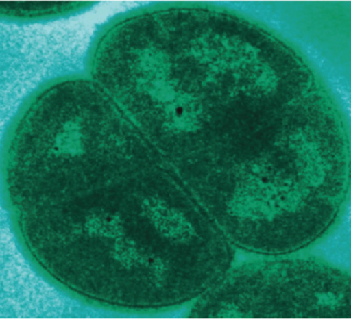
In high radiation environments, such as on the outside of the International Space Station or even on the surface of Mars today, some organisms, (e.g., Figure 13) have developed the ability to repair damaged DNA. You will learn later about the role of radiation in mitigating against contamination and why this microbial ability presents a challenge!
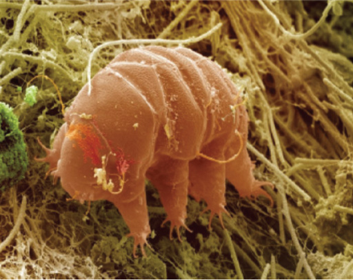
In the face of dry, desiccating conditions, some organisms (e.g., some bacteria, fungi, and even plants and animals such as Figure 14) survive by entering a state of apparent suspended animation where biological activity pauses. In the absence of water these organisms appear dead, but after days, weeks, or even years, if moisture returns, they reanimate and resume biological activities.
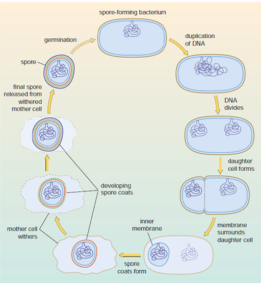
In some instances, a microbe’s only way of surviving extreme environmental conditions is to shut down most of its life processes and form spores. Spores are cells that have a multi-layered protective coating allowing them to exist, dormant, within an environment cannot be otherwise tolerated. Figure 15 shows the process by which spores form to protect the organism.
This protective coating can allow microbes to survive extreme heat, radiation and drought. Not all microbes can form spores, but the ability to form spores is found in many different species of bacteria and fungi.
Microbial spores are very common in the air around us and settle out onto surfaces both indoors and outdoors. They have even been found preserved in ancient tombs for several thousands of years. If conditions in the environment improve (e.g., the cell detects nutrients, water and less extreme conditions), then the spore can germinate, turning back into a non-dormant and un-coated cell that can replicate and grow.
The hardy nature of spores, combined with their ability to survive for thousands of years and still change back into a growing cell, makes them a major focus for planetary protection and clean room sterilisation.