

2.4 Obesity and brain reward systems
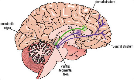
Very palatable foods, especially those high in fat and carbohydrate may be potent stimuli for neural pathways in the brain. Direct evidence for this idea has been found in recent studies of non-human animals. For example, it is known that many types of both natural (i.e. food, sex) and drug (e.g. cocaine or amphetamine, opiates, nicotine) rewards are able to stimulate activity within a brain pathway that innervates the ventral striatum at the base of the forebrain (Figure 8). The nerve cells that make up this pathway, which is called the mesolimbic dopamine projection, use dopamine as their neurotransmitter. Thus, when a rat eats a highly palatable and very sweet breakfast cereal there is an almost immediate increase in the release of dopamine within the ventral striatum. Other foods high in fat and carbohydrate are known to activate the same brain system.
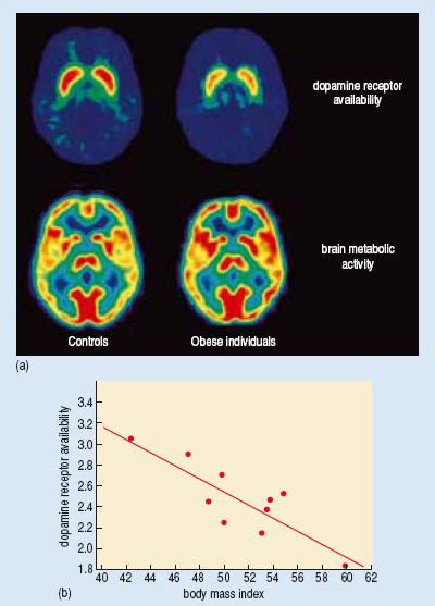
There is preliminary evidence that the same pathway may be involved in human responses to food-related cues. Recently Nora Volkow and her colleagues (Wang et al., 2001) used brain imaging techniques to visualize dopamine receptor availability in the brains of both normal weight and morbidly obese individuals (Figure 9a). They found significantly lowered dopamine receptor availability in the ventral striatum of the obese individuals.
Activity 24a
If dopamine receptor availability is lowered, what might be the effect on the functioning of the mesolimbic dopamine projection?
Answer
When the mesolimbic dopamine projection is stimulated to release dopamine, the cells in the ventral striatum will receive a smaller signal than usual because they have fewer receptors available to ‘pick up’ the neurotransmitters.
Activity 24b
The lower of the two sets of images in Figure 9a shows brain metabolic activity in obese and in control individuals. Why was it important to measure brain metabolic activity in this study?
Answer
The reduced dopamine receptor availability might have been a reflection of a much more general change in brain function. The absence of any difference in brain metabolic activity suggests that the change in dopamine receptor availability is a quite specific effect.
Activity 25
Use Figure 9b to describe the relationship between dopamine receptor availability and the degree of obesity.
Answer
The extent to which dopamine receptor availability was reduced was directly proportional to the degree of obesity; the more obese the individuals, the fewer the numbers of receptors available.
Volkow and her colleagues reasoned that the lowered dopamine receptor availability might generate pathological increases in eating as a compensatory response. Interestingly, similar decreases in dopamine receptor availability have been observed in the brains of people addicted to cocaine. However, in both cases, it is not possible to say whether the lowered dopamine receptor availability was the original cause of a pathological increase in eating, or an adaptive response that occurred only at a later stage.
There is also good evidence for the involvement of cortical areas of the brain in our response to the particular sensory characteristics of fat. Rolls and his colleagues (Rolls, 2004), working at Oxford, have used an imaging technique (fMRI) to demonstrate activation of parts of the frontal lobe, including the orbitofrontal and insular cortex, by fat-like stimuli. These two areas receive sensory inputs from the mouth and tongue and also have important connections to the ventral striatum (Figure 8).