
1.1.2 Lower respiratory tract
The trachea divides into two branches called bronchi (Video 2). These serve the left and right lungs. Like the trachea, the walls of the bronchi contain cartilage, which prevents their collapse. Each main bronchus divides into smaller and smaller tubes, finally ending in terminal bronchioles. Bronchioles also contain cilia that help to keep the airways clean by moving mucus and particles from the lower respiratory tract up to the pharynx to be expelled.
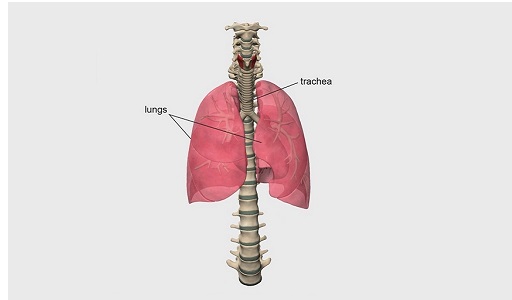
The lungs are organised into lobes (the left lung comprises two lobes and the right lung has three lobes). Two thin membranes, the visceral and parietal pleura, cover the lungs and keep them attached to the thoracic wall. The base of each lung is concave and rests on the diaphragm (more on that in Section 1.2), whereas the heart sits within the cardiac impressions, or grooves, in each lung (Figure 1, repeated).
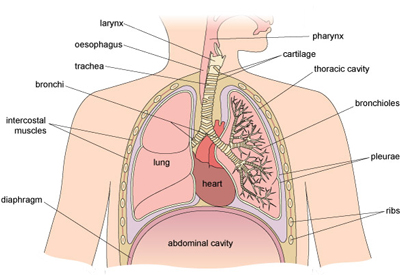
Because they act as a conduit for air to move into and out of the lungs, the nasal passages, pharynx, larynx, bronchi and bronchioles are collectively referred to as the conduction zone of the respiratory system.
Air then passes into progressively smaller structures deep in the lungs where gas exchange actually takes place in the respiratory zone, which you will explore in the next section.