

3.2 The anatomy of the language system
Perhaps the best-known generalisation about the language system is that it is represented on one side of the brain – usually the left – more than the other. Many lines of evidence support this view. Specific impairments to linguistic abilities are known as aphasia, and aphasia results much more often from damage to the left hemisphere of the brain than from damage to the right. It is also possible to temporarily deactivate one or other hemisphere. This is usually done as an investigative prelude to brain surgery, particularly in cases of epilepsy. It can be done by injecting a fast-acting drug into the carotid artery on one side of the body, or alternatively by weak electrical stimulation of part of the cortex. Deactivation of the left hemisphere causes a disruption of speech and language much more often than does deactivation of the right hemisphere. These techniques have also led to estimates that language is processed predominantly in the left hemisphere in about 97 per cent of right-handed people but in about only 60 per cent of left-handers.
We argued in the previous section that the initial perception of speech, like that of other sounds, was carried out bilaterally, whereas in this section we have argued that language is usually lateralised to the left hemisphere. There must thus be a point in the analysis of the speech signal where mechanisms on just one side ‘take over’ from the bilateral auditory areas. Brain scanning studies show that this is indeed the case: the additional activation in response to speech which is understood, compared to speech which is in an unknown language, is mainly on the left.
Further insight into such lateralisation comes from ‘split-brain’ patients. These are people who, because of severe epilepsy, have had the corpus callosum, the bundle of fibres connecting the two hemispheres, cut. This means that there is no direct neural connection between the left and right hemispheres. In these individuals, stimuli presented to the right hand, right ear, and right side of the visual field will mainly be processed by the left hemisphere, and stimuli presented to the left-hand side are processed by the right hemisphere. Objects presented to the left hemisphere can be named and talked about, whereas those presented to the right hemisphere generally cannot.
The right hemisphere does have some linguistic abilities. In split-brain patients the left hand can reach out and choose an object whose name has been presented to the right hemisphere, just as the right hand can choose an object whose name has been presented to the left hemisphere. However, the right hand can also choose the drawing that illustrates a sentence like The girl kisses the boy, whereas the left hand cannot correctly discriminate this situation from one in which the boy kisses the girl. Similarly, the right hemisphere cannot identify the scenes represented by The dog jumps over the fence versus The dogs jump over the fence, and so on. Thus, right-hemisphere language ability is limited to the phonological analysis of individual words, and access to their concrete meanings; sentence processing is the pure province of the left.
The areas within the left hemisphere which are most important for speech and language have been identified by several lines of evidence. Post-mortem examination of the brains of people affected by aphasia identified two main areas which were most often damaged – one in the frontal lobe and one further back, in the temporal lobe, close to the junction of temporal and parietal lobes. These areas are known respectively as Broca's area and Wernicke's area after the scientists who first identified their associations with language. Because of their respective positions, they are sometimes also called the anterior and posterior language areas. Evidence from non-aphasic individuals generally supports the importance of these areas. In one procedure, the cerebral cortex can be weakly electrically stimulated as a prelude to brain surgery for epilepsy. The patient can be awake during this procedure – since the brain contains no nociceptive neurons, only local anaesthesia is required. Thus, the patient can perform a verbal task whilst the electrical stimulation is moved around different areas of the brain.
Electrical stimulation disrupts speech and language activities mainly when it is on the left of the brain, and only in specific areas (Figure 15). These areas are in the general regions of Broca's and Wernicke's areas, as predicted. There is also an additional area in the left superior frontal lobe. This is in part of the cortex generally associated with motor functions, and may be important for the execution of speech production more than for language processing per se. There is also evidence for the involvement of tissues further down into the temporal lobe than the extension of the classical Wernicke's area/posterior language area. (Subcortical structures appear to be important, too, but they are beyond the scope of this course.)
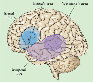
Brain imaging studies support the map shown above in Figure 15. Tasks to do with speech and language lead to increased regional blood flow – which is suggestive of greater neuronal ‘work’ – across a broad range of structures in the left hemisphere, generally within the triangle demarcated by Broca's and Wernicke's areas and the horn of the temporal lobe. There is strong evidence, which we will review in the next three sections, that this large area does not operate as an undifferentiated whole in the processing of language. Nor should we expect it to; as we saw in Section 2, decoding a sentence involves several distinct subtasks. The key questions to occupy researchers have thus been which subpart of the language cortex is associated with which subtask of overall language processing, and how does the processing of a sentence proceed in time through the various subareas and subtasks? The main lines of evidence we have to answer these questions come from three sources: aphasic patients, brain scanning and electrophysiological studies. We will look at each of these in turn in the next three sections.