
1.2 Muscles of ventilation
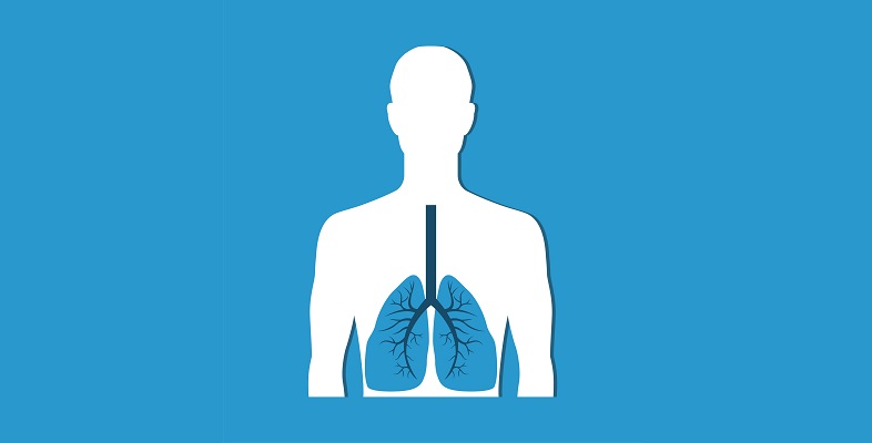
The expansion and contraction of the lungs is controlled mechanically by the diaphragm and the intercostal muscles (Figure 1, repeated).
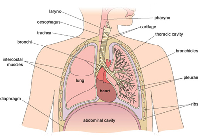
- The diaphragm is a dome-shaped muscle that sits underneath the lungs and separates the thoracic cavity from the abdominal cavity. It is innervated by the phrenic nerve, which originates in the medullary respiratory centre in the medulla of the brain.
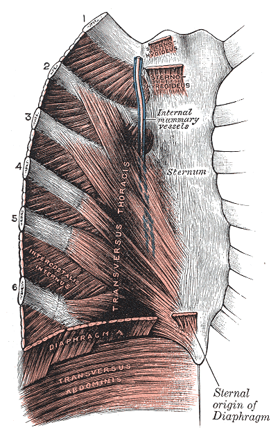
- The intercostal muscles are located in the ribcage. They receive neuronal inputs from intercostal nerves that arise from the thoracic nerves of the spinal cord.
The bronchi and bronchioles are also surrounded by smooth muscle cells that contract and dilate to regulate the amount of air that passes down to the alveoli.
Activity 2 Diaphragm and intercostal muscles
Part 1
To explore the location and function of the diaphragm and intercostal muscles, take a look at the anatomical information and diagrams on this site (open the links in a new tab/window so you can easily return to this page):
- Diaphragm [Tip: hold Ctrl and click a link to open it in a new tab. (Hide tip)]
- Intercostal muscles
Part 2
Now try matching each of these statements with the correct muscles.
Using the following two lists, match each numbered item with the correct letter.
-
flatten(s) when contracted to expand the size of the thoracic cavity and decrease the size of the abdominal cavity, generating a pressure gradient within the thoracic cavity
-
draw(s) the ribs upwards and outwards; most active during inhalation
-
draw(s) the ribs downwards and inwards; most active during forced exhalation
-
stiffen(s) the chest wall during respiration; most active during forced exhalation
a.external intercostal muscles
b.innermost intercostal muscles
c.internal intercostal muscles
d.diaphragm
- 1 = d
- 2 = a
- 3 = c
- 4 = b