
2.2.4 Film cassette and grid
As the X-rays pass through the patient some of them will be scattered and will therefore not follow the expected line through the patient. If these reach the detector they will blur the image. Some of the scattered radiation can be removed by a grid, usually oscillating, placed between the patient and the detector.
Analogue imaging systems use either film alone (rarely) or a combination of a film and fluorescent material (phosphor). The phosphor fluoresces and produces visible light which is recorded by the film. The film is then developed.
Digital imaging systems use a variety of methods to record the intensity for each pixel in the image. This can then be displayed on a VDU or printed out on film.
Activity 2
See if you can answer the following questions. Please think about your answer before clicking to reveal the answer.
What happens to most of the electrons which strike the anode in the X-ray source, and approximately what percentage of electrons produce X-rays?
Answer
Most of the electrons produce heat, only 1 per cent of the electrons produce X-rays.
Activity 3
Why is a grid place in front of the film?
Answer
The grid reduces the amount of scattered X-rays which reach the film, thus producing a clearer or sharper image.
Activity 4
Can you think of some advantages and disadvantages of X-ray imaging?
Answer
The advantages of X-ray imaging are:
X-ray images are relatively cheap to produce;
images can be acquired quickly and are particularly useful for a rapid initial assessment;
X-ray images are readily accessible.
The disadvantages are:
ionizing radiation dose;
2-D image of a 3-D object;
contrast can be poor.
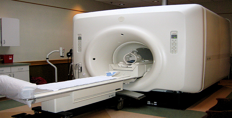
The disadvantage of planar X-ray imaging, producing a two dimensional representation of a three dimensional object, is overcome by computed tomography (CT) imaging.