
6.4 Taking the image
Activity 14
Now watch this video clip of a patients lungs being imaged, called a VQ (ventilation quotient) scan. What are the two different types of acquisitions used called? What radioactive substance is used for each acquisition, and why?
Click to view clip about planar scans [2 minutes 21 seconds]
Transcript: Planar scans
Answer
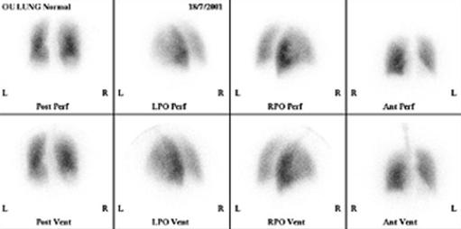
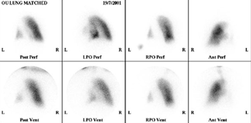
The two different scans mentioned were a perfusion and ventilation scan, to look at the blood supply and air supply to the lungs, respectively. In order to do this simultaneously two different radionuclides that have different gamma ray energies are used. This is a valuable diagnostic test for a pulmonary embolism (a blood clot in the lungs).
To image the perfusion the patient is given an injection of technetium-99m (gamma energy 140 keV, half-life 6 hours) combined with macro-aggregated albumin (MAA). The ‘large’ particles of MAA tend to lodge in the very small capillaries of the lungs and therefore give a good indication of where there is, or is not, a good blood supply.
The ventilation can be imaged by asking the patient to breathe a mixture of air and krypton-81. This is produced from a generator containing the parent radionuclide, rubidium-81. As the patient is breathing the krypton-81, it shows which areas of the lungs the gas reaches, and therefore, which areas are ventilated.
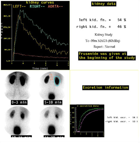
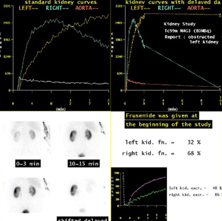
Another widely used procedure is a dynamic renogram. In this case the radionuclide imaged is one that is taken up and excreted by the kidneys. Successive images are collected over a period of time (e.g. every 15 seconds for 20 minutes) and can then be analysed to compare the function of the kidneys.
Activity 15
Now watch this final video clip on radionuclide images, on the single photon emission computed tomography (SPECT) imaging technique. The sequence shows cardiac (heart) images being obtained. How does SPECT produce slices through the body?
Click to view clip about SPECT [3 minutes 23 seconds]
Transcript: SPECT
Answer
Data is collected from a large number of directions by rotating one or more camera heads around the body. Then, tomographic images can be reconstructed, by filtered back projection, using a similar process to CT. The speed of rotation is much slower than that used in CT – the whole process taking at least 10 minutes.
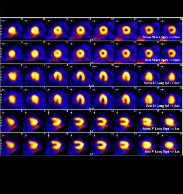
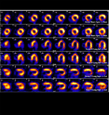
Images of a normally perfused heart muscle (myocardium), and a heart with an inferior and lateral wall defect are shown below.
Activity 16
Take a moment to consider what you think the advantages and disadvantages of radionuclide imaging would be compared with ultrasound, MRI, CT and X-rays.
Answer
The advantages of radionuclide scans are:
demonstration of functional information that often cannot be obtained in other ways;
wide variety of organs can be imaged;
tomographic and 3-D images available (SPECT).
The disadvantages are:
poor resolution;
radiation dose to the patient;
slow and labour intensive;
specialised radiopharmacy and scanners are not readily available at all hospitals.