
5 Ultrasound
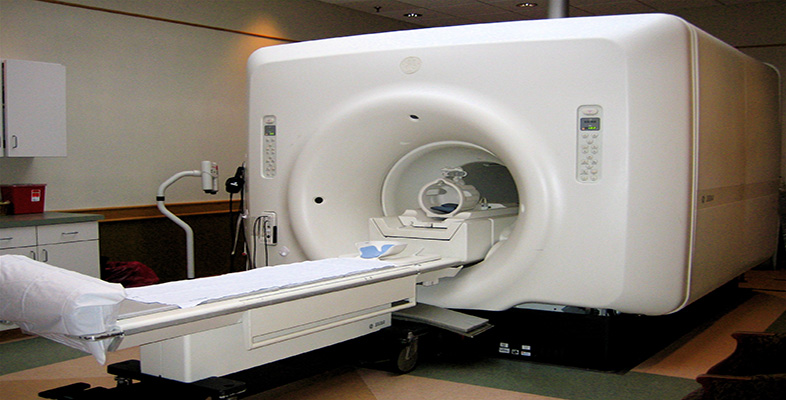
Ultrasound imaging uses acoustic waves, rather than ionizing radiation, to form an image. The principle is rather like radar; a pulse of ultrasound (1–15 MHz) is sent out from the transducer and reflected from tissue boundaries. Measurement of the time taken for the pulse to return allows the distance to the reflecting boundary to be calculated.
The important parameter determining the amount of reflection is known as the acoustic impedance (Z) of the tissue and is the product of acoustic velocity and density.
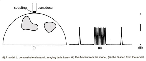
The appearance of the returning echoes can be displayed in two principal ways. Firstly the amplitude of the echo can be shown as a vertical displacement against a horizontal time axis, which takes the appearance of a profile such as that typical of a mountain range. This is known as A (amplitude) mode.
The alternative is to show the echo intensity as dots of varying brightness and this is known as B (brightness) mode. 2D imaging uses a large number of adjacent B-mode lines to form the final image, a B-scan.
In a third mode, called M (movement) mode, a single line of a B-scan is chosen and the position of the reflecting boundaries is plotted as a function of time. A moving boundary will show up as a wavy trace. The shape of this trace can be indicative of critical features such as the operation of cardiac valves.
Activity 10
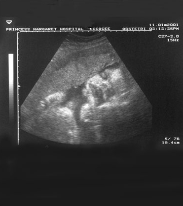
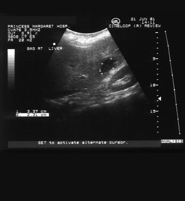
Look at these ultrasound images of a fetus, and the liver and kidneys. Do you think it was obtained using A-mode, B-mode or M-mode ultrasound imaging?
Answer
They are both B-mode images. This is the type of ultrasound image we are all most familiar with.
Activity 11
Watch the following video clip.
Click to view clip about ultrasound [5 minutes 24 seconds]
Transcript: Ultrasound
What advantages of ultrasound imaging are mentioned?
In addition to B-mode imaging, which other forms of ultrasound imaging are shown?
Answer
Ultrasound imaging is quick, cost effective, has virtually no known hazards and is acceptable to most patients.
M-mode, colour flow Doppler and flow velocity/time image.
We will now look in a little more detail at the ultrasound transducer. Design of transducers is complex but they rely on the piezo-electric effect. When a voltage is applied across piezo-electric materials they change shape. If an a.c. voltage is used then the crystal vibrates at the same frequency and sound is produced.
The process also works in reverse – sound incident on the crystal gives rise to an a.c. voltage. Thus the same crystal can be used to transmit and receive.
Each transducer will be designed for use at a particular frequency. In general a higher frequency gives better resolution but lower penetration. So for large patients, or deep structures, the frequency will have to be low – perhaps 3 MHz; for small structures, such as the eye, frequencies can be much higher (e.g. 12 MHz). Some transducers are designed to transmit at one frequency and receive at another, allowing detection of the second harmonic reflections.
Transducers can be designed for use externally or via body orifices such as the vagina (for uterine imaging), or the oesophagus (for heart imaging).