
1 Current imaging techniques
A grainy Polaroid of the child in a mother's womb – an X-ray of a tibia fractured in a traffic accident – a report on a brain scan anxiously awaited. Very few of us have not had some connection with the techniques and practices of medical imaging. Often, these contacts are in periods of personal drama in which the medical images chart our physical status, the management of a condition and, in some cases, our future.
Imaging is a central feature of contemporary medicine. Along with chemically based techniques of analysis, it provides the clinician with ways of extending investigations that go beyond external observation and interview.
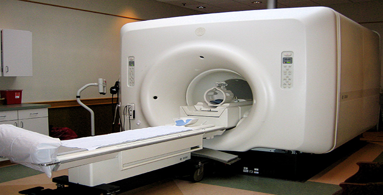
The video clip below will introduce the major imaging modalities in current use in hospitals. These are:
X-rays, including computed tomography (CT)
ultrasound
magnetic resonance imaging (MRI)
radionuclide imaging
The view offered in the clip is that of the underlying science, from the physical scientist's perspective, rather than that of the medical clinician.
Activity 1
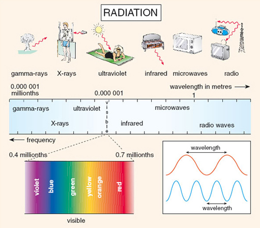
As you watch the video clip, keep in mind the electro-magnetic spectrum shown in Figure 1 below and try to place the electromagnetic energy of each modality on the spectrum.
Click to view the introduction clip presented by Liz Parvin [7 minutes 50 seconds]
Transcript: Introduction clip presented by Liz Parvin
Answer
X-rays and CT use X-ray part of the electromagnetic spectrum.
Medical ultrasound uses sound waves rather than electromagnetic radiation. The frequency of the sound waves is usually between 1 and 15MHz.
Magnetic resonance images uses radio waves (usually around 50–150MHz).
Radionuclide imaging (not actually shown in the video clip, but mentioned by Alan Davis) uses gamma radiation.