
6.3.1 Collimator
Without a collimator, gamma rays from all directions would be collected by the crystal and no useful image could be obtained. Gamma rays cannot be focused by a lens but a collimator consisting of a series of holes in a lead plate can be used to select the direction of the rays falling on the crystal. Most collimators in use today are parallel hole collimators. A parallel hole collimator is shown schematically in Figure 16.
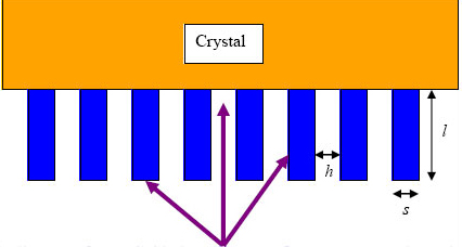
Figure 16: Schematic diagram of a parallel hole collimator. Gamma rays are shown in purple. Obliquely incident gamma rays are absorbed in the collimator septa
The resolution and sensitivity of a collimator depend on a number of factors including:
hole size (h);
the thickness of the septa (s), the lead between the holes;
the length of the holes (l);
the energy of the gamma rays.
Different collimators are selected for different procedures – e.g. Low Energy High Resolution.