
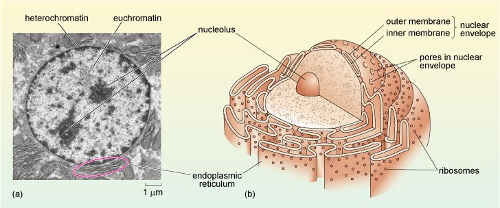
Figure 11 (a) Electron micrograph of the nucleus of a rat liver cell. Heterochromatin and euchromatin, which are described in the text, can be clearly differentiated as electron-dense (dark) and electron-lucent (pale) areas, respectively. Two electron-dense nucleoli (see below) are also clearly visible. (b) Schematic diagram showing the structure of the nucleus, which is linked to the endoplasmic reticulum.