

1 Introducing the cell
Studies into the function and appearance of cells reveal the presence of great diversity, which is particularly striking among eukaryotic cells. Even very different types of eukaryotic cells, however, exhibit many common structures and functions. This mix of uniformity and diversity is also reflected in the organisation within cells. This course looks in more detail at the interior of cells - their subcellular structure, including their 'ultrastructural' features (sometimes called 'fine structure'), which are visible using electron microscopy. Knowledge of the subcellular components of cells and how these components are arranged is fundamental to your understanding of how cells perform their functions: that is, how cells 'work'.
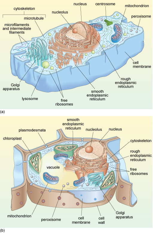
Schematic diagrams of typical animal and plant cells are shown in Figure 1a and b. You do not need to study this figure in detail now; it will be referred to again during the course of this course.
Before beginning your study of cell components, some of the main techniques used to study the interior organisation of cells are briefly outlined.