6.4 Rapid-response genes and rhythmic neuronal activity
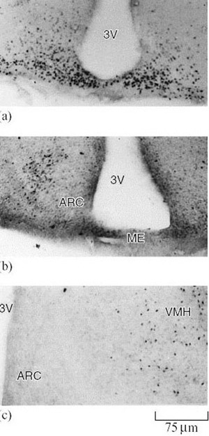
Reactive changes in the brain are usually marked by changes in neuronal electrical activity. If these changes are to be of long duration, adjustments in neuronal electrical behaviour may be made through changes in gene expression. Rapid-response genes (sometimes called ‘immediate-early’ genes) are activated within minutes of the onset of such sustained electrical activity. These genes are master controls, acting as a gateway to a series of linked events: alteration of electrical firing patterns, repetitive activity and even the structure of neurons and their synapses, all of which modify the output of specific functional groups of nerve cells. Two such genes, called c-fos and junB, whose expression pattern in the SON follows the circadian rhythm in euthermic jerboas (Jaculus orientalis), cease cyclical fluctuations and remain at constant, elevated levels in the hypothalamus during bouts of hibernation. The region containing the largest number of c-fos-positive neurons in the hibernating jerboa is called the arcuate nucleus (Figure 40a). As hibernation progresses, and then on arousal, the distribution of c-fos-positive cells shifts to the ventromedial hypothalamus (Figures 4.39b and c). The significance of this observation is that separate regions of the brain are involved in maintenance of the hibernating state and initiation of locomotor activity on arousal (Ouezzani et al., 1999).

Question 18
Where else might you expect to see c-fos-positive cells in the brain on emergence from hibernation?
Answer
In regions involved in initiating motor activity to the muscles.
Since the expression of both c-fos and junB genes changes in advance of alterations in neuronal behaviour, they are likely to act as signals for the re-emergence of euthermic electrical activity patterns on arousal. It is significant that the high levels of expression are located in the arcuate nucleus, separate from the SON, which could therefore be an independent regulator of locomotor activity. During hibernation, the lowest temperature for electrical activity of hypothalamic neurons is depressed from around 16° C to 12.3° C and there is an increase in the number of cold-sensitive neurons. In the European hamster (Cricetus cricetus), neurons in isolated slices of the hypothalamus can fire quite normally down to temperatures as low as 5° C as a result of changes in the molecular configuration of ion channels in the cell membrane. Such an adaptation is particularly important because neural control systems must be intact at brain temperatures that prevail on entry to, and during arousal from, hibernation. The structure of neuronal dendrites (see Figure 41a) is also modified to reduce incoming synaptic contacts. All three changes result from subtle alterations in the expression of genes specific to neurons.
