3 Anatomical imaging using MRI
The potential of nuclear magnetic resonance (NMR) spectroscopy to be applied to investigations of the human body was recognised soon after the technique was developed in the 1940s. But its use as an imaging technique to visualise anatomy was first shown to be practicable in the 1970s.
Since then, magnetic resonance imaging (MRI) has become an astonishingly common procedure.
For example, figures released by the National Health Service in England reveal there were 2.4 million MRI examinations carried out during the financial year 2012/13.
The following quotation from MRI from Picture to Proton (Cambridge University Press) illustrates both the complexity and the utility of the technique:
MRI involves an amazing combination of advanced science and engineering, including the use of superconductivity, cryogenics, quantum physics, digital and computer technology – and all within the radiology department of your local hospital (Robbie et al., 2007).
-
Can you suggest why MRI is potentially less harmful to patients than CT?
-
Unlike X‑ray-based diagnostics such as CT, MRI does not expose patients to potentially harmful ionising radiation.
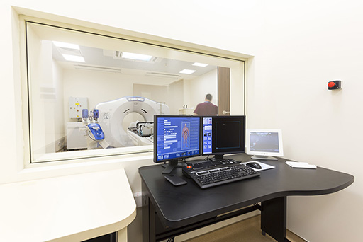
A typical MRI scanner is shown in Figure 5.