2.5 Intracellular receptors
Signal receptors are usually located at the cell surface. However, it is important to remember that there are some groups of receptors that do not fit into the general signal transduction model set out in Figure 2, These are intracellular receptors, which bind small or lipophilic molecules such as steroid hormones, which can cross the cell membrane. The signalling pathways activated by these receptors seem quite simple compared with the other pathways we shall be dealing with, but the same principles of ligand binding, conformational change, signal amplification, translocation and so on described earlier still apply.
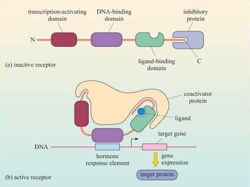
One important family of intracellular receptors are the nuclear receptors (also known as ‘nuclear hormone receptors’), which includes receptors for steroid hormones, thyroid hormones, retinoids and vitamin D. Although the ligands differ in their structural type, all nuclear receptors are structurally similar. They are good examples of receptors with intrinsic transcriptional activity (Section 1.3), comprising a transcription-activating domain, a DNA-binding domain and a ligand-binding domain. Their ligands are all small and hydrophobic, and so they can diffuse readily through the plasma membrane. The receptors are usually held in an inactive conformation by inhibitory proteins (often chaperones/heat-shock proteins). Binding of the ligand induces a conformational change that causes the inhibitory protein to dissociate from the receptor (Figure 27). The receptor may then translocate to the nucleus if it was in the cytoplasm, or it may already be in the nucleus; either way, the receptor–ligand complex is now able to bind to specific DNA sequences by means of its DNA-binding domain. Binding to DNA can also be facilitated by association of the receptor–ligand complex with other proteins (referred to as ‘coactivator proteins’). The DNA sequence to which the receptor–ligand complex binds is a promoter region of the target genes; in the case of hormones, it is called a ‘hormone response element (HRE)’.