

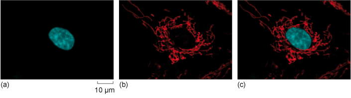
Figure 11 HeLa cells labelled with two fluorescent dyes: one that binds to DNA and one that accumulates inside mitochondria. (a) Micrograph taken after illuminating the cells with ultraviolet (UV) light to excite the dye bound to DNA, which then emits blue light. (b) A second micrograph taken after illuminating the cells with green light to excite the dye localised in mitochondria, which then emits red light. (c) The images are merged using computer software to show the fluorescence of both dyes at the same time.