

2.2 Fluorescence (light) microscopy
A particularly insightful technique that scientists use to obtain information about the structure and function of the inner workings of cells is by using fluorescent light for visualiation. Here, we don’t include the term ‘light’ in the name and just call it fluorescence microscopy (also called fluorescence imaging).
Fluorescence is very much part of the natural world. You may be aware of fluorescent lighting, perhaps in your home or workplace, but fluorescence is also abundant in minerals, plants and animals. The jellyfish Aequorea victoria is an example of an animal that uses fluorescence as part of its defence against predators (Figure 6).
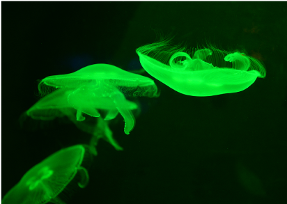
This jellyfish became famous when the gene that encodes the protein that makes it appear fluorescent, green fluorescent protein (GFP), was isolated and cloned. GFP has since been used extensively in cell biology research, along with many other fluorescent proteins that have different colours.
The awarding of the 2008 Nobel Prize in Chemistry to Roger Tsien, Osamu Shimomura, and Martin Chalfie for their work in characterising green fluorescent protein (GFP) highlights the profound impact of fluorescent proteins on biological research. Their groundbreaking contributions have revolutionised how scientists visualise and study cellular processes in real time. (For more information on GFP and its applications, see the ‘Further reading’ section at the end of this course.)