

2.3 Components of a fluorescence microscope
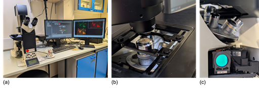
Fluorescence microscopes are similar to conventional light microscopes but include special filters that allow specific wavelengths of light to illuminate the sample and detect the emitted fluorescence (Figure 7). While understanding the technical details of these filters is not necessary for this course, it is helpful to know that they enable the detection of fluorescent signals from labelled cell and tissue components. When working with living cells growing in a dish or flask, researchers often use a so-called inverted microscope. In an inverted microscope, the objectives are located below the sample and the light is directed from above, making it easier for the researcher to access and manipulate the sample.
Like conventional light microscopy, fluorescence microscopy can be used in living or fixed cells. There are several methods for staining specific structures and proteins within cells for visualisation using fluorescence microscopy. Because the structures are viewed against a dark background, fluorescence microscopy provides much greater contrast, making it easier to clearly identify and distinguish cellular structures.
Why might you want to use an inverted microscope and have access to the sample when working with living cells?
Answer
You might want to exchange the medium that is covering the cells to ensure they are kept alive, or in order to add compounds to stimulate the cells so you can observe their response in real time.