

2 Types of microscopy
The three different microscopy techniques most commonly used to study cells are light microscopy, fluorescence (light) microscopy and electron microscopy. The first two use light to visualise the sample, while electron microscopy uses a beam of electrons. Because cells have a very limited contrast, they are difficult to see clearly under any microscope without staining. Various staining techniques can be used to visualise structures, or even specific proteins, inside cells. The choice of staining method depends on the type of microscopy, and the particular research question being addressed.
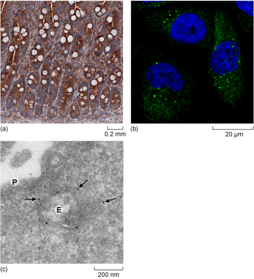
Each type of microscopy has its own advantages and limitations, which you will explore as you learn more about the three types. Examples of cells visualised with each technique are shown in Figure 4.
What difference can you observe in the scale bars shown in Figure 3?
Answer
The scale bars range from 0.2 mm to 200 nm, indicating that the images taken with a light microscope reveal fewer details than those taken with fluorescence (light) microscopy. However, they allow us to see the whole cells.
Which of the panels in Figure 3 was taken with the highest resolution?
Answer
The image taken with a transmission electron microscope has the highest resolution and reveals most details.
Box 2 Use of scale bars in microscopy
The magnification of a structure in a micrograph, an image taken using a microscope, results as a combination of several steps in which the original structure is magnified. First, the microscope objective applies a magnification. Then the camera used for taking the image can apply an additional magnification. Finally, the computer software displaying the image can also apply magnification or display the image in various sizes, depending on your screen. That’s why stating the magnification is not the most useful way to show the size of a structure, and where the use of scale bars is critical.
A scale bar is added to a micrograph to illustrate the size of the cells or structure independent of the equipment used to display it, and independent of the size of the image that is displayed. It can be shown within or underneath the micrograph. In multi-panel figures, one scale bar might apply to several images.
You will now be introduced to the components of light microscopes.