

5.2 Fluorescence microscopy allows visualising dynamic processes
Fluorescence microscopy is a powerful tool for observing dynamic processes like the movement of mitochondria inside cells (Video 2).
While you might picture mitochondria as many small, oval-shaped structures scattered throughout the cell, fluorescence imaging reveals that in many cases, they actually form an interconnected, network-like structure. This network is often in motion, constantly changing shape and position. As a result, the simplified appearance of mitochondria in static micrographs doesn’t fully reflect their complex and dynamic nature.
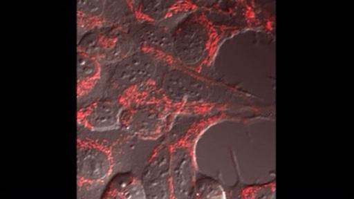
Transcript: Video 2 Mitochondrial dynamics. Mitochondria in HeLa cells expressing a red fluorescent protein were visualised using fluorescence microscopy. Constant movement of mitochondria can be seen in all cells. The cell towards the bottom left shows changes in the shape of its elongated mitochondria.
Why is it not possible to obtain a video like Video 2, showing mitochondrial movement, when using electron microscopy?
Answer
To be used in electron microscopy, the cells need to be fixed. They are no longer alive and no movement will take place inside the cells.
Visualising these dynamic processes greatly helps with understanding how a cell functions. Comparing how such processes are affected, for example in the context of a disease, can help to understand how diseases develop and affect cellular behaviour.