

4.4 Expression of fluorescent proteins
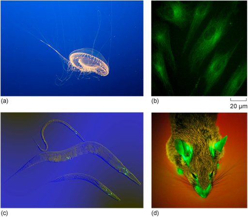
Some dyes can be used in living cells, but one of the most powerful and widely used labelling techniques in live cultured cells, and even whole organisms, is to genetically modify them to express a fluorescent protein. Interestingly, certain animals naturally produce fluorescent proteins as part of their defence against predators. One well-known example is the jellyfish Aequorea victoria, which produces GFP, a molecule that you have encountered earlier in the course (Figure 13a).
Scientists can take the gene that encodes GFP and fuse it to a gene of interest and then introduce this modified gene into cells (Figure 13b) or even whole animals (Figure 13c and d). The resulting GFP-fusion protein can be visualised in real time under a fluorescence microscope, allowing researchers to track where the protein is located inside the living cell. In most cases, the fusion protein behaves like the original protein, maintaining its normal function and proper localisation within the cell.
You have now learned about the different types of microscopy, the principles of fluorescence and how cellular structures using fluorescence. In the next section you will briefly explores why fluorescence microscopy is considered such a versatile technique in cell biology.