

5.1 Fluorescence microscopy allows observing specific structures individually and combined
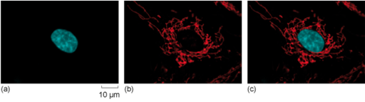
Fluorescence microscopy makes it possible to observe specific cellular structures both individually and in combination. After staining different structures within a cell using fluorophores of different colours, the microscope’s separate channels can be used to select specific wavelengths of light to excite each dye one at a time. This allows for the capture of individual images, each showing a single labelled structure (Figure 14a and b, and Video 1). These individual fluorescence images are then digitally merged or overlaid using computer software, creating a combined image that reveals the spatial relationship between all stained structures within the cell or tissue (Figure 14c).
Capturing separate images and merging them allows detailed studies of the localisation of structures and proteins. You will see more examples when using the digital fluorescence microscope yourself.