

4.1 Journey into a cell
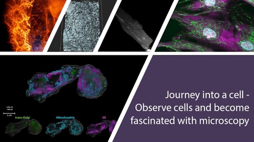
Video 1 provides you with a ‘Journey into a cell’, which uses images and videos to illustrate how cell structures and organelles can be viewed within living cells, and how multiple fluorescent probes can be used within the same cell or tissue to build high-resolution two- and three-dimensional representations. The following sections will explain some of the techniques mentioned in the video in more detail.
Watch the video and then answer the following questions.
Transcript: Video 1 ‘Journey into a cell’
Which technique allows scientists to mark and visualise structures inside living cells?
Answer
Fluorescence microscopy (not electron microscopy).
How can you identify the nucleus in a cell?
Answer
By applying a DNA-binding probe to visualise it by its fluorescence, or by the space it occupies being left ‘dark’ when probes are used that detect other cellular organelles that are present in the cytoplasm.
Can you think of a reason why using more than three colours in fluorescence microscopy might prove difficult?
Answer
It can be difficult to distinguish the colours/organelles stained.
What conclusions can you draw from experiments done with the fluorescent staining of living cells and tissues that you would not be able to draw using fixed material (where the cells/organs were killed before staining)?
Answer
The ability to use living cells and tissues means that you can study cells in their native environment and also examine interactions between cells and how they change over time. You can also observe the movement of organelles and/or molecules inside cells in real time.
What example was given in Video 1 of fluorescence being used to measure the change in concentration of an ion over time?
Answer
Calcium ion changes in the cardiac myocyte.
From your ‘Journey into a cell’, you should now have a good understanding of how fluorescence can be used to study cells and tissues. Some of the techniques mentioned in the video will be explained in more detail in the following sections.
You will now learn about some techniques that are used to stain structures and proteins inside cells before they can be inspected using fluorescence microscopy. You will see examples of these staining techniques later when you inspect cells in the digital fluorescence microscope.