

2.4 Electron microscopy
Although it’s not the focus of this course, electron microscopy was mentioned because of its higher resolution and ability to study ultrastructure in detail. You will now be briefly introduced to two important types of electron microscope (EM).
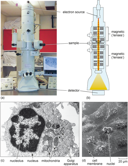
Transmission electron microscopy: a beam of electrons is accelerated in a transmission electron microscope (TEM) at high velocity through a sample, which is a very thin section (less than 10 nm). Samples can range from tissue sections to purified protein complexes. Electrons cannot pass through glass; instead, magnets are used as the ‘lenses’ that control and focus the path of the electron beam. The interior of the microscope is under vacuum to prevent scattering of the electrons by air molecules. Electrons that have passed through the sample reach the detector, where they activate a fluorescent screen, or are captured with a digital camera, forming the image (Figure 8a and b). Electron microscopes first became available in 1939 and assisted in the discovery of organelles like the Golgi apparatus (Figure 8c).
Scanning electron microscopy: a technique for studying the surface of intact cells using a scanning electron microscope (SEM). The sample is first coated with a thin metallic layer that deflects an electron beam onto the detector, giving a very fine detail of the surface features (Figure 8d).
Electron microscopy can only be used in fixed cells. During the sample preparation, heavy metals are used to increase the contrast in the samples, for example to clearly see cellular membranes.