

2.1 Components of a light microscope
All microscopes use a series of magnifying lenses to enable very small objects such as cells to be seen by the human eye. A light microscope (or optical microscope) uses visible light to illuminate the sample and glass lenses to focus and magnify the image. Light microscopy can magnify an object up to about 1000 times the original size, obtain a resolution of around 200 nm and it can be used with either living or fixed cells. The box below briefly explains how a light microscope works.
Box 3 How a light microscope works
In a light microscope, three lenses are important for forming the magnified image: the condenser, the objective and the camera (Figure 5).
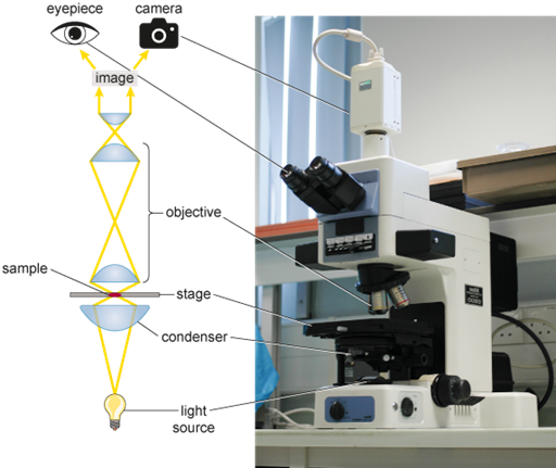
The condenser focuses a beam of light onto the sample placed on the stage. In an upright microscope, like the one shown in Figure 5, the light source and condenser are both located beneath the stage. The focused beam of light is transmitted through the sample and then passes through the objective, which magnifies the image and passes it to the eyepiece(s), or to a camera that directs the captured image to a computer screen. The image is brought into sharp focus by moving the sample closer to, or further from, the objective. Most microscopes have several objectives ranging from ×4 to ×100 magnification (note that ‘×’ means times and indicates the times-fold magnification). The eyepiece adds further magnification, often ×10. For example, the total magnification achieved by a ×4 objective combined with a ×10 eyepiece will be 4 × 10 = ×40. Often, additional magnification is added by the camera taking the images, or within computer software.
To increase the contrast and visualise specific structures in cells or tissues, different staining techniques can be used. Light microscopy is very important as a technique to detect changes in cell structure during the development of diseases and are routinely used in pathology labs around the world. If you are interested to learn more about histology, the study of the microscopic structure of complex plant and animal tissues, the free OpenLearn course ‘Histology, microscopy, anatomy and disease’ provides a lot more information. In this course, you will now take a look at fluorescence microscopy.