5.3 Specialised applications: Confocal microscopy
Specialised applications of fluorescence microscopy are continuously being developed, and the examples presented here present only a snapshot – this list will undoubtedly expand in the future. While this course does not cover these advanced techniques in detail, they are introduced to give you an idea of the wide-ranging possibilities fluorescence microscopy offers.
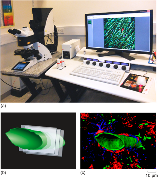
Confocal microscopy (Figure 15a) enhances image clarity by eliminating out-of-focus light, using a pinhole and laser scanning system. This allows for the capture of a series of optical sections – thin, focused slices – at different depths within a specimen. These images form a z-stack (Figure 15b), which computer software can reconstruct into a highly detailed three-dimensional (3D) image of the cell or tissue (Figure 15c). This is particularly useful for studying complex structures in thick specimens, such as tissues or organoids, and provides insights into spatial relationships within cells.