

1.1 Why do we need microscopy?
Almost all cells are too small to be seen with the naked eye, so the study of cellular structure only began with the development of lenses and microscopes that could magnify cells many hundreds of times. A typical bacterium, for example, is about one micrometre in diameter and no more than a few micrometres in length (Box 1).
Box 1 Units used to measure the size of cells
To get down to the scale of cells, a unit of length is needed that is one-thousandth of a millimetre. This unit is the micrometre – abbreviated to µm (µ is the Greek letter mu) and sometimes referred to as a micron.
An even smaller unit, called the nanometre (abbreviated to nm), is needed when describing the size of subcellular components such as organelles, the membrane-bound structures inside eukaryotic cells that have a specific function. A nanometre is one-thousandth of a micrometre (Table 1).
| Unit (symbol) | Multiple in metres | Multiple in micrometres |
|---|---|---|
| metre (m) | ||
| centimetre (cm) | ||
| millimetre (mm) | ||
| micrometre (µm) | ||
| nanometre (nm) |
If you are not familiar with the very small units of measurement mentioned in Box 1, you should study Figure 2 and work through the questions that follow. Familiarity with the relative sizes of various molecules and organisms will be helpful for any future study in this area.
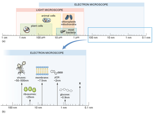
How many nanometres are there in one millimetre?
Answer
There are 1000 (103) nm in 1 µm and there are 1000 (103) micrometres in 1 mm. So, there are 1000 × 1000 = 1 000 000 (or 106) nm in 1 mm.
What type of scale is used in Figure 2 and why is it used?
Answer
It is called a logarithmic scale. Each unit is ten times greater than the previous unit, which is helpful when you want to show a very wide range of values using the same scale.
In Figure 2, approximately what size is the structure labelled as X?
Answer
Just under 0.3 nm, which is the size of a water molecule.
What is the size range (from smallest to largest) of bacteria in micrometres and in nanometres?
Answer
0.8–10 µm, which is 800 nm to 10,000 nm (or to ).
Next, you’ll be introduced to the important difference between magnification and resolution, which helps you to understand why some microscopy techniques allow you to see more detail.