

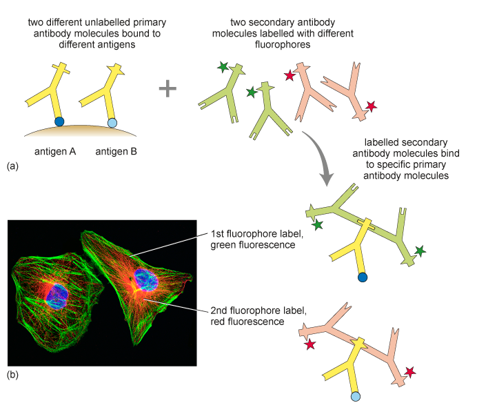
Figure 12 Double immunolabelling of the cytoskeleton. (a) Illustration of the principle of double immunolabelling using two primary antibodies and two differently labelled ‘secondary’ antibodies. Note that the epitopes on the primary antibodies, to which the secondary antibodies bind, differ in shape. (b) Double immunofluorescence staining in cells using a primary antibody against actin (the secondary antibody was labelled with a green fluorophore) and tubulin (the secondary antibody was labelled with a red fluorophore). The nucleus is shown in blue.