
My OpenLearn Profile
Personalise your OpenLearn profile, save your favourite content and get recognition for your learning
Exploring cells with digital fluorescence microscopy
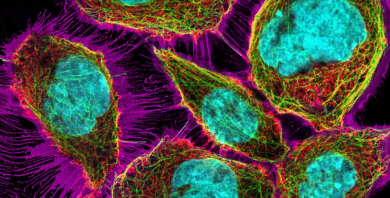
Learn about microscopy techniques, understand their differences and delve into the fascinating world of fluorescence microscopy. You’ll learn how to make proteins and structures fluorescent, and you’ll see why fluorescence microscopy is one of the most versatile techniques in cell biology. You can refresh your knowledge about cellular structures using a 3D interactive cell. After seeing many fascinating examples of the use of fluorescence microscopy, you’ll use a digital fluorescence microscope to explore samples yourself.
Course learning outcomes
After studying this course, you should be able to:
- outline some of the key principles of microscopy (with a focus on fluorescent microscopy) and the differences between light, fluorescence (light) and electron microscopy
- describe some labelling techniques used in fluorescence microscopy
- explore the structures and organelles found in a cell via fluorescence microscopy
- understand how to use a digital fluorescence microscope to collect data and images
- interpret images and data collected using fluorescence microscopy.
First Published: 31/03/2026
Updated: 31/03/2026
Rate and Review
Create an account to get more
Track your progress
Review and track your learning through your OpenLearn Profile.
Statement of Participation
On completion of a course you will earn a Statement of Participation.
Access all course activities
Take course quizzes and access all learning.
Review the course
When you have finished a course leave a review and tell others what you think.
Access to the OU's online microscope: "https://students.open.ac.uk/stem/openlearn/microscope/ver/1.1.4/?dataset=s296%5Cs296_fluo" was clearly extremely beneficial in gaining what approached to hands-on experience (without the hassle of creating samples!)
The comparative and scenario based review questions were very useful, though the final "Quiz" less so.
Miscellaneous notes:
It came as a surprise to see the 6 "Editor" mark-ups that were present in the MS Word version; the "Introduction" being a case in point, with 3.
Introduction
"[Editor: course image to be added (banner on front page / top of each page)]
[Editor: I thought one of the DFM images would be a logical pick. I couldn't find any of those specific images on the protein atlas site (to find the largest / highest res version), so I've dropped in something else that felt visually striking... any thoughts on that, or something else specific we should use?]
[Editor: source for image currently in place, HPA056283: HeLa: https://www.proteinatlas.org/ENSG00000114745-GORASP1/subcellular]"
3.1 "[Author: SAQ to be added?] [Editor: - not sure if this was still to be decided? all fine either way, we can drop something in here, or I can just remove this comment if not needed]"
4.5 Table 2 stated that Transmission electron microscopy (TEM) achieves up to ~1,000,000x, whilst the following states potentially beyond TEM 50,000,000 magnification?
https://www.nanoscience.com/blogs/whats-the-difference-between-sem-and-tem/
Electron microscopy labelling: "Limited; specific structures identified by morphology" I was not clear whether this covered Immunogold labelling?
https://en.wikipedia.org/wiki/Immunogold_labelling
5.2 Mitochondrial dynamics
I am not sure that the video did "*clearly* show how the mitochondria move and change their shape."
5.2 The online course post-video 2 question is absent from the MS Word document...
"Why is it not possible to obtain a video like Video 2, showing mitochondrial movement, when using electron microscopy?"
...though the question has been covered before anyway.
5.5 Specialised applications: Super-resolution microscopy
The following URL provides one update on nm resolution, whilst including some informative graphics...
https://www.hamamatsu.com/eu/en/applications/life-sciences/fluorescence-imaging/super-resolution-microscopy.html
Once again a question is absent from the MS Word document...
Which cellular structure with a diameter of ~25 nm can you think of, that you could visualise with super-resolution microscopy?
6 Inspecting structures in a 3D cell
cytosTolic -> cytosolic
Question 1 the MS Word list is already in the correct order.
7 Using the digital fluorescence microscope
Refers to "...your Practical Workbook" ...in this context presumably this is intended to refer to the course itself?
"Now go to the DFM in [Editor: add live link]"
https://students-dev.open.ac.uk/stem/openlearn/microscope/ver/1.1.4/?dataset=s296%5Cs296_fluo
...is present, rather than...
https://students.open.ac.uk/stem/openlearn/microscope/ver/1.1.4/?dataset=s296%5Cs296_fluo
8 Quiz
Question 6 "[Editor: double check first sentence here – both answers were listed as dyes but I think the second should’ve been indicators? (I’ve adjusted accordingly)]"